Can Anomaly Scan Be Done at 19 Weeks? A Practical Guide
Explore whether a fetal anomaly scan at 19 weeks is appropriate, what it reveals, and tips to optimize image quality within the 18–22 week window. Scanner Check analyzes timing, expectations, and practical steps.

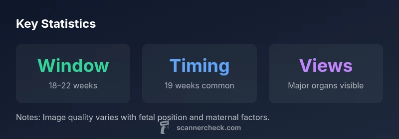
Yes. According to Scanner Check, an anomaly (fetal anatomy) scan at 19 weeks is within the standard window of 18–22 weeks gestation. Most providers can obtain a thorough assessment of fetal anatomy, heart, limbs, spine, and organ development at this stage, assuming favorable fetal position and no maternal or medical factors hindering the exam.
Anatomy scan timing and the 19-week window
The fetal anatomy, or anomaly, scan is a detailed ultrasound designed to evaluate major organ systems and skeletal structures. The conventional window for a comprehensive anatomy survey is 18–22 weeks gestation. At 19 weeks, many pregnancies yield excellent visualization of the brain, spinal column, heart, liver, kidneys, stomach, and limb buds. This timing balances the maturation of fetal organs with the likelihood that anatomy is still clearly delineated on ultrasound. Providers may schedule within this window for convenience, fetal growth considerations, or prior risk factors. When the scan falls at 19 weeks, expect a systematic check of all major structures and a careful assessment of placental position, amniotic fluid, and overall fetal growth. According to Scanner Check, 19 weeks is a well-supported timing for reliable findings that influence prenatal care decisions.
What the anomaly scan evaluates at 19 weeks
During a 19-week anatomy scan, clinicians systematically assess: cranial structures (brain ventricles, skull shape), facial features (where views permit), spinal alignment, thoracic organs (heart chambers, great vessels, diaphragm), abdominal organs (liver, stomach, kidneys), and limb development. The goal is to identify major congenital anomalies and to confirm expected growth patterns. While the 19-week window is suitable for most fetuses, certain conditions or maternal factors may alter image quality or reveal findings more clearly at a slightly earlier or later time. Communication with your care team helps tailor the timing to your pregnancy.
Factors that influence image quality at 19 weeks
Image clarity at 19 weeks depends on multiple factors. Fetal position is a primary determinant; a fetus facing away from the transducer or with limbs obscuring key views can complicate assessment. Maternal body habitus, abdominal wall thickness, and the amount of amniotic fluid also affect image quality. Ongoing medical conditions, placenta previa, or scarring can complicate visualization. In some cases, providers may recommend rescheduling or performing targeted views to ensure critical structures are evaluated. Preparation and a calm environment can help, but position is often the deciding factor for a diagnostic-quality exam.
When 19 weeks might not yield ideal views
Even within the 18–22 week window, suboptimal views can occur. If significant anatomy cannot be assessed adequately, clinicians may propose a repeat scan within a short interval or plan a follow-up evaluation at a later gestational age (commonly 20–24 weeks). The decision hinges on the specific body systems being evaluated, prior risk factors, and the likelihood that a clearer image will be obtainable later. In such cases, patients are usually advised about benefits and limitations of repeating the study and what findings would trigger further testing.
How clinicians handle suboptimal views and follow-ups
When views are incomplete, clinicians may perform targeted measurements, adjust the imaging approach, or schedule a repeat scan to confirm anatomy. In cases where a potential anomaly is suspected, a referral to a maternal-fetal medicine specialist might be made for enhanced assessment and counseling. Documentation typically includes the structures visible, any findings, and recommendations for follow-up imaging. Clear communication with the family about the window for additional views helps manage expectations and reduces anxiety.
Preparing for the ultrasound and questions to ask
Before the scan, review your pregnancy history and any risk factors with your clinician. Bring a list of questions such as: Which organs will be emphasized in this exam? If some views are not possible now, when should we expect a follow-up? How will findings be communicated, and what would trigger additional testing? Hydration guidance varies by practice; some centers prefer a full bladder for transabdominal scans in early pregnancy, while others do not require it. Wearing comfortable clothing and arriving a little early can ease the process.
Real-world scenarios: what to expect after the scan
Many 19-week scans yield reassuring results, with confirmed anatomy and normal growth trajectories. If a minor finding is noted, clinicians may schedule a confirmatory scan or additional tests. In rare cases, more extensive testing, including genetic counseling or fetal echocardiography, may be recommended based on findings and parental preferences. Regardless of the outcome, you will receive documentation detailing what was observed, what was not assessable, and the plan for any follow-up imaging.
Anatomy scan timing window overview
| Aspect | Typical Timing | Notes |
|---|---|---|
| Window | 18–22 weeks | Standard anatomy scan window |
| 19 weeks timing | Within window | Often yields clear views if fetus is favorable |
| Follow-up possibility | If views are incomplete | May schedule 1–2 weeks later or at 20–24 weeks |
Common Questions
Is 19 weeks too early for the anomaly scan?
No. 19 weeks falls within the standard 18–22 week window for a thorough anatomy scan, and many clinics conduct detailed views at this time.
No—19 weeks is within the normal window for a detailed anatomy scan.
What if the baby isn’t cooperating at 19 weeks?
If the baby isn’t in a good position, technicians may retry the exam later or schedule a follow-up scan to obtain the necessary views.
If the baby isn’t in a good position, you may need a follow-up scan.
Can an anomaly scan miss problems at 19 weeks?
Subtle findings can be harder to see early; most major structures are visible, but some details may be clearer later in pregnancy.
Most major structures are seen, though some details can be clearer later.
What factors affect scan quality at 19 weeks?
Fetal position, maternal body habitus, amniotic fluid level, and placenta location can influence visibility.
Body habitus and fetal position often affect image clarity.
Should I schedule the scan now or wait?
Discuss with your clinician. The 18–22 week window is standard, and 19 weeks is a common, well-supported choice.
Most people schedule within 18–22 weeks; 19 weeks is fine.
What should I ask the technician during the scan?
Ask what was seen, whether all major structures were evaluated, and if any findings require follow-up, plus the plan for next steps.
Ask what was seen and whether follow-up is needed.
“The anatomy scan is most informative when performed between 18 and 22 weeks; at 19 weeks, clinicians typically see major structures clearly and can provide actionable findings.”
Key Takeaways
- Schedule within the 18–22 week window for anatomy visualization.
- Fetal position heavily influences image clarity; be prepared for potential rescan.
- Ask about follow-up scans if any views are incomplete or ambiguous.
- Discuss risk factors with your clinician to tailor timing and expectations.
- Most major structures are assessable by 19 weeks, with options if needed.