Can You Get a Scan at 3 Weeks Pregnant? What to Expect
Explore whether you can get a scan at 3 weeks pregnant, what early imaging can reveal, and how to confirm pregnancy with tests and timing guidance from Scanner Check.

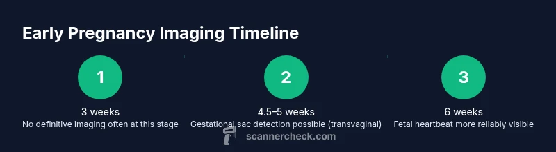
Can you get a scan at 3 weeks pregnant? Generally no. Most providers cannot visualize a pregnancy on ultrasound at 3 weeks gestation. Transvaginal ultrasound may detect a gestational sac around 4.5–5 weeks, with fetal heartbeat around 6 weeks. In early pregnancy, doctors rely on pregnancy tests (urine or blood hCG) and clinical symptoms to confirm pregnancy and dating. Always consult your obstetrician or midwife for personalized guidance.
Understanding 3 weeks into pregnancy
For someone asking can you get a scan at 3 weeks pregnant, it helps to ground expectations in what gestational age means. At this very early stage, a pregnancy may still be too small to visualize on ultrasound. The uterus, fallopian tubes, and early gestational products are typically not yet detectable by standard imaging. This is also why many clinicians rely on biochemical and clinical markers rather than imaging alone at this point. According to Scanner Check, the timing of imaging depends on gestational age, symptoms, and medical history; the body’s hormonal signals and subjective symptoms often guide the plan as much as imaging does. Understanding this nuance helps you plan the next steps with your care team.
Earliest imaging timelines: what can you see and when
Ultrasound visibility in very early pregnancy is a moving target. A transvaginal ultrasound, which places the probe closer to reproductive organs, may begin to show a gestational sac around 4.5–5 weeks gestation for many people. A fetal pole or visible heartbeat is typically detectable closer to 5.5–6 weeks, though every person’s timeline can vary. If you are at 3 weeks, it is common for the scan to appear normal or show nothing definitive. This timeline is influenced by factors such as maternal BMI, pregnancy location (uterus vs. ectopic risk), and the equipment’s sensitivity. The Scanner Check analysis highlights that imaging in the very early weeks is more about ruling out complications and confirming dating rather than providing a definitive pregnancy map.
How pregnancy is confirmed before ultrasound: the role of hCG
Because a 3-week scan is unlikely to yield clear findings, clinicians frequently rely on human chorionic gonadotropin (hCG) levels to confirm an early pregnancy. Blood tests can provide a precise estimate of pregnancy progression and help with dating when ultrasound timing is uncertain. A rising hCG level typically indicates a viable early pregnancy, but the rate of rise varies. Home urine pregnancy tests are convenient for initial detection, but blood tests offer quantitative data that can guide later imaging decisions. The key idea is that imaging and lab tests complement each other in the first weeks of gestation.
When to seek medical care for pain or bleeding in early pregnancy
If you experience heavy bleeding, severe pain, dizziness, or fainting at any stage of early pregnancy, seek urgent care. While light spotting can be common, heavy bleeding or cramps can signal an issue that merits immediate evaluation. Your healthcare provider will determine whether imaging or additional tests are needed based on symptoms, history, and risk factors. Early evaluation can help diagnose ectopic pregnancy, miscarriage risk, or other concerns. Remember, this is a time when you may not see much on ultrasound, so timely clinical assessment matters more than the image alone.
What to ask your clinician about early scans and dating
If an early scan is on the table, prepare questions to maximize clarity. Ask about the earliest gestational age detectable for you specifically, what findings would be expected at each stage, and how imaging will influence your care plan. Inquire about the benefits and limitations of early ultrasound versus relying on hCG data. Understanding the plan helps you feel more confident and reduces unnecessary worry while you wait for the next milestone.
Preparing for an appointment: practical steps
Before your visit, gather dates of your last period, any prior pregnancy history, and a list of symptoms. Bring results from home pregnancy tests and any orders from other providers. Write down questions in advance to ensure you cover your priorities during the appointment. Having a clear plan reduces stress and helps your clinician tailor the right testing strategy for your situation.
Safety, accuracy, and limits of imaging in early pregnancy
Ultrasound is considered a safe, noninvasive imaging modality. In very early pregnancy, its accuracy depends on the gestational stage and equipment sensitivity. The limits of detection mean you might not see a clear gestational sac at 3 weeks, even if pregnancy is present. Your provider will balance ultrasound results with lab tests and clinical assessment. If imaging timelines shift, don’t panic—care teams adjust plans based on how your pregnancy progresses across weeks.
Alternatives and next steps if initial imaging is inconclusive
If an initial scan around 3 weeks yields unclear results, clinicians typically plan a follow-up scan around 6–7 weeks when visualization improves dramatically. In the meantime, rely on hCG monitoring and clinical guidance to track pregnancy viability. If symptoms worsen or new concerns arise, contact your healthcare provider promptly for reassessment and a tailored imaging schedule.
How to track your pregnancy timeline and plan follow-up imaging
Maintaining a simple timeline can help you stay organized. Track last menstrual period, estimated due date, and any pregnancy test dates. If your doctor recommends a repeat ultrasound, mark it on your calendar and set reminders. A structured plan reduces anxiety and ensures you don’t miss critical milestones as pregnancy progresses.
Tools & Materials
- Prenatal appointment with obstetrician/midwife(Essential for guidance on early imaging and dating strategy)
- hCG blood test(Provides quantitative confirmation and helps with dating before imaging)
- Home pregnancy tests(Convenient initial detection; bring results to appointment)
- Access to ultrasound scheduling(Needed to plan imaging windows and follow-ups)
Steps
Estimated time: 1-2 weeks (depends on scheduling and clinical needs)
- 1
Schedule your initial prenatal appointment
Call or use the patient portal to book a visit with your obstetrician, midwife, or primary care provider. The goal is to discuss early pregnancy imaging timing, confirm next steps, and establish a plan for follow-up testing if needed.
Tip: Bring your last menstrual period date and any positive home test results to help with dating. - 2
Review dates and symptoms with the clinician
During the visit, share the expected gestational age, any pain or bleeding, and previous pregnancy history. This helps the clinician decide whether an early ultrasound is warranted or if lab tests should take precedence.
Tip: Ask specifically when an ultrasound would be most informative given your dates. - 3
Ask about earliest imaging options and what can be seen
Discuss the likelihood of visualizing a gestational sac at your current gestational age and what findings would indicate a typical progression. If imaging is pursued, clarify what the results would mean for your care plan.
Tip: Request a clear plan for next steps if imaging is inconclusive. - 4
Request non-imaging confirmation if appropriate
If imaging is not immediately informative, discuss quantitative hCG monitoring and other laboratory tests to confirm pregnancy and help with dating before repeat imaging.
Tip: Note any signs that would trigger an earlier revisit, such as pain or heavy bleeding. - 5
Plan follow-up imaging around 6–7 weeks if needed
If pregnancy continues, schedule a follow-up ultrasound when visualization is more reliable. This helps confirm viability and assess fetal development milestones.
Tip: Set a reminder and ask what preliminary signs clinicians look for at this stage.
Common Questions
Can you detect pregnancy at 3 weeks with ultrasound?
Usually not; 3 weeks is typically too early to visualize a gestational sac. Imaging often becomes informative around 4.5–5 weeks for a sac, and around 6 weeks for a heartbeat.
Usually not; three weeks is too early to visualize a pregnancy on ultrasound. Most findings become visible around five to six weeks.
When is the earliest ultrasound for pregnancy?
The earliest reliable ultrasound is commonly around 4.5–5 weeks for a gestational sac with a transvaginal exam; heartbeat is typically seen around 6 weeks.
The earliest ultrasound is usually around 4.5 to 5 weeks for a sac, with a heartbeat seen around six weeks.
What tests confirm pregnancy before ultrasound?
Urine or blood hCG tests can confirm pregnancy before imaging and help with dating when ultrasound timing is uncertain.
Pregnancy tests, especially blood tests measuring hCG, can confirm pregnancy before an ultrasound.
Is an early scan safe?
Ultrasound is considered safe when performed by trained professionals; early scans are done when clinically indicated and follow medical guidance.
Yes, ultrasound is safe when done by professionals, and early scans are used when needed.
What should I bring to my appointment?
Bring dates of your last period, any home test results, and a list of symptoms to help the clinician tailor testing.
Bring your last menstrual period date, test results, and any symptoms to your appointment.
Can a scan replace a pregnancy test?
No. Imaging does not replace the need for pregnancy tests, and both imaging and lab data are used together to guide care.
No—imaging doesn't replace pregnancy tests; doctors use both together to guide care.
Watch Video
Key Takeaways
- Expect no visible signs on ultrasound at 3 weeks.
- Rely on hCG testing for early confirmation and dating.
- Plan follow-up ultrasound around 6–8 weeks if pregnancy continues.
- Ask your clinician about timing and options for early imaging.