Does DEXA Scan Have Radiation? Safety, Doses, and What to Expect
Learn whether a DEXA scan involves radiation, how the exposure compares to daily background levels, safety considerations for pregnancy, and what to expect during the procedure.

does dexa scan have radiation? In short, yes, but the exposure is extremely small. A DEXA scan uses two low-dose X-ray beams to measure bone density, and the overall radiation dose is tiny compared with common imaging tests. For most adults, the benefit of accurate bone health assessment outweighs the minimal risk. If you’re pregnant or have concerns, talk with your clinician.
What a DEXA Scan Measures and How It Works
A DEXA (dual-energy X-ray absorptiometry) scan is a specialized imaging test designed to assess bone mineral density (BMD). It targets key areas such as the hip and spine to determine fracture risk and diagnose osteoporosis or osteopenia. The scan uses two X-ray beams at different energy levels. By comparing how each beam is absorbed as it passes through bone and soft tissue, the machine computes a precise BMD value. The process is quick and noninvasive: you lie on a padded table while a sensor passes smoothly over the area to be measured. Modern scanners are calibrated to minimize dose while maximizing accuracy. In many clinics, the technician will guide you to breathe normally and stay still for a few seconds to ensure a clean image. This test is widely considered essential for monitoring bone health, especially in populations at risk for osteoporosis.
does dexa scan have radiation
does dexa scan have radiation? The short answer is yes, but the dose is exceptionally small. DEXA relies on low-dose X-ray beams, and the total exposure for a typical scan is far below the levels associated with more intensive imaging like CT. In practice, this means most adults receive a radiation dose that is considered negligible for routine bone health assessment. Shielding is not routinely required because it can interfere with the measurement, though clinicians assess each case individually, particularly if there are special considerations such as pregnancy. The overall risk from a single scan is typically described as minimal, and the benefits of accurate bone density data generally outweigh this tiny exposure. If you have specific concerns about radiation, discuss them with your healthcare provider before the appointment.
Comparing DEXA to Other Imaging Tests
DEXA is distinct from other imaging modalities in its purpose and exposure profile. Unlike CT, PET, or some fluoroscopic exams, a DEXA scan is designed to measure bone density and uses a very low, well-controlled radiation dose. A standard chest X-ray or CT scan delivers noticeably higher radiation than a DEXA scan, while MRI does not use ionizing radiation at all. When considering imaging for bone health, DEXA offers a targeted, low-dose option that provides clinically meaningful information with minimal exposure. This relative safety profile helps clinicians monitor conditions like osteoporosis over time, enabling treatment adjustments without subjecting patients to significant radiation.
Who Benefits Most from a DEXA Scan
DEXA scanning is particularly valuable for people at risk of osteoporosis or low bone density. This includes postmenopausal women, older adults, individuals with a family history of fractures, and patients on long-term corticosteroid therapy. Men and younger adults with certain conditions may also benefit when risk factors or symptoms warrant evaluation. Insurance guidelines and clinician judgment often determine frequency, but routine checks are common for those with known risk factors or prior fragility fractures. A DEXA scan can inform lifestyle changes, nutritional adjustments, or pharmaceutical interventions aimed at preserving bone health over time.
What to Expect During the Test and After
During a DEXA scan, you typically remove jewelry and metal clothing and lie flat on the scanner table. The technician positions your body to target the hip and/or spine and may ask you to hold still for a few seconds while images are captured. The actual scanning time is brief, and results are usually available within a short follow-up visit or a few days. Your radiologist or clinician will interpret the T-score and Z-score to assess bone density, compare with reference populations, and guide management. If results indicate low bone density, lifestyle changes, calcium/vitamin D optimization, or medications may be recommended. Follow-up scans are often scheduled to monitor changes over time.
Safety, Pregnancy, and Special Considerations
Pregnant individuals are advised to avoid nonessential radiation exposure when possible. If a DEXA scan is clinically indicated during pregnancy, clinicians carefully evaluate the risk-benefit ratio and may adjust the timing or use alternative assessments. For people with extreme obesity or metal implants, imaging may require adjustments in positioning or technique, but the goal remains to obtain accurate bone density data. Talk with your doctor about any implants, pregnancy plans, or prior imaging studies so the technologist can tailor the approach while maintaining safety and diagnostic quality. Scanner Check emphasizes patient-specific risk assessment to minimize unnecessary exposure while preserving diagnostic value.
Practical Tips for Preparation and Understanding Results
Plan your appointment with convenience in mind: wear loose clothing, avoid heavy metallic accessories, and bring any prior scan results for comparison. Understanding DEXA results requires reviewing the T-score and Z-score with your clinician; these metrics help categorize bone density relative to a young reference group and to your age-matched peers. Discuss potential lifestyle changes, vitamin D and calcium intake, and activity levels that support bone health. If repeat scans are recommended, ask about the interval and what to watch for in between exams. Scanner Check reminds readers to use the results as a guide alongside overall health status, not as a single diagnostic decision.
AUTHORITY SOURCES
- National Institutes of Health (NIH) Osteoporosis and Related Bone Diseases National Resource Center: https://www.bones.nih.gov
- U.S. Food and Drug Administration (FDA): Radiation-Emitting Products Guidance: https://www.fda.gov
- National Institute for Health and Care Excellence (NICE): Osteoporosis Guidelines: https://www.nice.org.uk
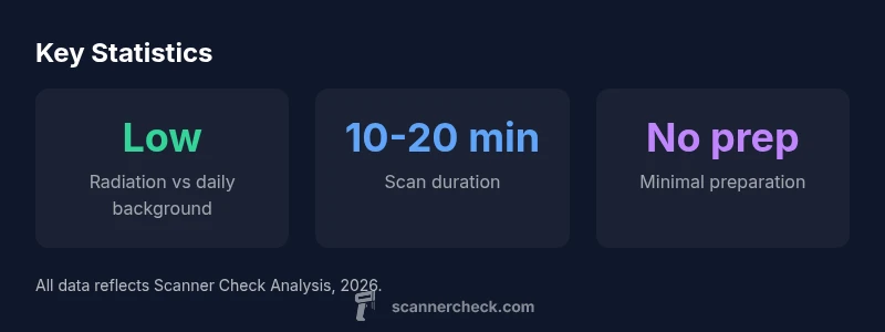
DEXA scan at a glance
| Aspect | Typical Characteristics | Notes |
|---|---|---|
| Radiation dose | Very low (qualitative) | Two low-dose beams; minimal exposure |
| Exam duration | about 10-20 minutes | Short procedure; positioning matters |
| Preparation | Minimal | Remove metal items; no fasting required |
Common Questions
Is the radiation from a DEXA scan harmful to most people?
For most patients, the radiation dose from a DEXA scan is very small and not associated with meaningful risk. Clinicians weigh benefits against exposure and typically recommend the test when bone health information will influence treatment.
The radiation is very small, and doctors usually recommend the test when it can change your care plan.
Can I have a DEXA scan while pregnant?
Pregnancy requires careful consideration. If bone density data is essential, your clinician may proceed with precautions or delay until after pregnancy. Always inform your imaging team about pregnancy status.
If you're pregnant, tell your clinician; they will weigh risks and may adjust timing.
How long does it take to get results?
Results are typically available within a few days to a week, depending on the facility. A clinician will review the T-score and Z-score to interpret bone density and guide next steps.
Results usually come back in a few days.
How often should I have a DEXA scan?
Frequency depends on initial results, risk factors, and treatment plans. Your clinician will determine an appropriate interval, often years apart for stable bone density.
Your doctor will tell you how often to repeat the test.
Are there alternatives to DEXA for bone health?
Other methods like quantitative ultrasound or serum bone turnover markers exist, but DEXA remains the standard for diagnosing osteoporosis and tracking changes over time.
There are alternatives, but DEXA is the standard for bone density.
What should I do before and after the test?
Wear comfortable clothing, avoid metal, and schedule follow-up discussions to review results with your clinician. Hydration and overall bone-healthy lifestyle contribute to better outcomes alongside imaging.
Wear comfy clothes, then discuss results with your doctor.
“The DEXA scan provides essential information about bone density with minimal radiation exposure, making it a trusted tool in osteoporosis risk assessment.”
Key Takeaways
- DEXA scans involve radiation, but exposure is very low.
- The test is widely used for osteoporosis risk assessment.
- Pregnant individuals should discuss timing with their clinician.
- Results guide treatment decisions and follow-up imaging cadences.