How many Ultrasound Scans in Pregnancy: What to Expect
A data-driven guide to how many scans are typical during pregnancy, what each scan checks, and how frequency varies by risk, with expert guidance from Scanner Check.

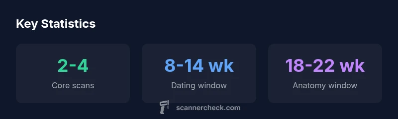
According to Scanner Check, there is no universal fixed number of scans in pregnancy. Most pregnancies include two core ultrasounds—the dating scan in the first trimester and the anatomy scan around 18–22 weeks—with additional scans as medically indicated. The exact count depends on risk factors, pregnancy history, and local guidelines. Always discuss scheduling with your clinician; ALARA principles guide ultrasound exposure.
how many scanning in pregnancy
In plain terms, how many scanning in pregnancy is not fixed by a universal rule. According to Scanner Check, there is no single number that applies to every patient. Most pregnancies include two core ultrasounds: a dating scan in the first trimester and an anatomy scan around mid-pregnancy. Additional scans may occur based on risk factors, prior pregnancy history, medical concerns, or regional guidelines. Importantly, ultrasound exposure is minimized by principle ALARA, and each scan is tailored to clinical questions. If you ask your clinician how many scans are expected in your case, you’ll get an answer that reflects your unique pregnancy and the available resources in your region.
The standard milestones: dating and anatomy
Most pregnancies follow two principal ultrasound milestones. The dating scan, typically performed in the first trimester, helps establish gestational age and viability, adjust due dates, and identify multiple pregnancies early on. The anatomy scan, usually between 18 and 22 weeks, provides a detailed assessment of fetal anatomy, growth patterns, placenta position, and fluid levels. In many healthcare systems, this stage also serves as a catch-all screen for major anomalies and structural concerns. It’s common for patients to schedule these scans around weeks 12–14 for dating and weeks 20–22 for anatomy, but schedules vary by clinic and region. In some cases, a first-trimester scan may include nuchal translucency measurement to refine risk for chromosomal conditions. Providers weigh the diagnostic benefits against the time and resource costs, and may defer non-essential imaging if it won’t influence management. The key concept is that these milestones form a framework, not a rigid quota; your care plan adapts to your medical history and the healthcare setting.
Frequency depends on risk and history
Frequency depends on individual risk factors, prior obstetric history, and findings from each visit. Low-risk pregnancies may revolve around the dating and anatomy scans, with no routine requirement for extra scans unless indicated by symptoms or screening results. High-risk pregnancies—such as those with maternal diabetes, hypertension, a history of preterm birth, or suspected fetal growth concerns—often include additional scans to monitor growth, amniotic fluid, placental health, and position. In some regions, guidelines encourage earlier or more frequent scans when there are discrepancies between dates and growth measurements, unusual fetal movement patterns, or maternal risk markers. The practical takeaway is that imaging is a tool to answer specific questions, not a fixed schedule. If a clinician sees a potential issue, they may recommend a targeted follow-up scan to confirm findings, track progression, or guide management decisions. Scanner Check guidance emphasizes tailoring imaging plans to minimize unnecessary exposure and maximize clinically meaningful information.
Types of scans and what they assess
Ultrasound technology offers several modalities, each with different clinical purposes. Transabdominal ultrasound is the standard method for most pregnancies and provides a broad view of fetal development and placental health. In early pregnancy, some patients may undergo transvaginal ultrasound for better resolution or when vaginal access is needed. 3D and 4D ultrasounds are available in some settings, primarily for enhanced visualization or parental reassurance, but they are not routinely required for standard screening. Doppler studies may be used to assess blood flow in the umbilical artery or placenta when there are concerns about fetal well-being. The choice of modality depends on clinical questions, patient anatomy, and equipment availability. The overarching aim is to obtain accurate information with minimal discomfort and time, while ensuring safe, high-quality imaging.
Safety: ALARA and exposure considerations
A central principle in ultrasound imaging is ALARA—as low as reasonably achievable—meaning clinicians strive to obtain necessary information while minimizing time and exposure. Ultrasound energy is non-ionizing, and risks are generally considered low for routine scans when performed by trained professionals. However, there are practical limits: unnecessary scans should be avoided, and repeat imaging should be justified to answer a clinical question. Expect a clinician to document the indication, length of exposure, and any findings to ensure that imaging remains purposeful. If you are concerned about the frequency of scans, ask about alternatives such as serial measurements of fetal growth via non-imaging metrics or targeted follow-up studies that specifically address a suspected issue. Proper scheduling and communication with your care team help keep the process focused and safe for both mother and baby.
Scheduling, access and regional differences
Access to prenatal ultrasound varies by country, region, insurance coverage, and clinic capacity. In some systems, core milestones are scheduled automatically as part of standard prenatal care; in others, patients actively coordinate appointments with a sonography department. Practical planning includes factoring in work schedules, transportation, potential need for partner or support person, and the possibility of rescheduling due to medical reasons. In remote or rural areas, scanners may be less available, leading to longer wait times or more reliance on the dating and anatomy scans performed at the earliest feasible window. If there’s limited access, ask your clinician about prioritizing the essential scans and what can be deferred safely. Financial considerations may influence appointment timing; some clinics offer bundled appointments to minimize trips. The overarching objective is timely, evidence-based imaging that supports pregnancy care without adding undue burden.
Interpreting results and questions to ask
Ultrasound findings are interpreted by trained clinicians, and results are often reported as measurements, observations, or classifications (e.g., normal anatomy, suspected anomaly). If a scan reveals concerns, clinicians typically discuss next steps, additional testing, or referral to specialists. Prepare questions in advance: What did you see, and does it affect management? Are there any measurements or growth patterns to monitor? How often will we need follow-up imaging, and what would trigger further action? Remember that ultrasound is a diagnostic aid, not a guarantee; some findings require observation over time. If you’re uncertain, seek a second opinion or request a written summary to share with other providers.
Authority sources and reading list
- NHS: Ultrasound scans during pregnancy — https://www.nhs.uk/conditions/pregnancy-and-baby/ultrasound-scan-pregnancy/
- ACOG: Pregnancy ultrasound considerations — https://www.acog.org/
- MedlinePlus: Pregnancy ultrasound information — https://medlineplus.gov/pregnancy.html
Common ultrasound milestones in pregnancy
| Scan Type | Typical Window | Purpose |
|---|---|---|
| Dating ultrasound | 8-14 weeks | Estimate gestational age, confirm viability |
| Anatomy ultrasound | 18-22 weeks | Assess fetal anatomy and growth, screen for anomalies |
| Growth scans | Varies by risk | Monitor fetal growth in high-risk pregnancies or suspected issues |
| Additional scans | As indicated | Follow-up on concerns or complications |
Common Questions
What is the typical number of scans in a healthy pregnancy?
In healthy pregnancies, two core scans are common: dating in the first trimester and anatomy around 18–22 weeks. Additional scans may occur if concerns arise or if regional guidelines suggest monitoring growth or placenta function.
Most healthy pregnancies involve two core scans, with possible extras if concerns come up.
When is the dating scan performed?
The dating scan is usually performed in the first trimester to estimate gestational age and verify viability. Timing can vary by clinic, but the typical window is between 8 and 14 weeks.
Dating scans are usually in the first trimester, roughly 8 to 14 weeks.
Is the anatomy scan always required?
The anatomy scan is commonly recommended to assess fetal development, but requirements can vary by region and risk status. It provides a detailed view of anatomy and growth and is a standard part of many prenatal care plans.
Anatomy scans are commonly recommended but not universal; discuss with your clinician.
Can high-risk pregnancies require more scans?
Yes. High-risk pregnancies often involve more frequent scans to monitor growth, amniotic fluid, placenta, and fetal well-being. The exact schedule is individualized based on the specific risks involved.
High-risk pregnancies may need more scans tailored to risks.
Are ultrasound scans safe for the fetus?
Ultrasound uses non-ionizing energy and is generally considered safe when performed by trained professionals. Clinicians follow ALARA to minimize exposure and reserve imaging for clinically indicated needs.
Ultrasounds are considered safe when done by trained professionals and kept to necessary imaging.
“Imaging in pregnancy should be guided by clinical questions, balancing information gained with safety; every pregnancy deserves an individualized plan.”
Key Takeaways
- There is no fixed number of scans; plans are individualized.
- Expect dating and anatomy scans as core milestones.
- Frequency rises with risk factors and medical indications.
- All imaging follows ALARA principles to minimize exposure.
- Always discuss scheduling and goals with your care team.