How Often Can You Have Scans: A Practical Imaging Frequency Guide
Learn how often you can have scans, what drives imaging frequency, and how to minimize exposure while preserving diagnostic value. Practical guidance from Scanner Check on CT, MRI, X-ray, and ultrasound.

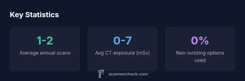
How often can you have scans depends on medical need, scan type, and safety considerations. There is no universal cap, but providers aim to minimize radiation exposure. For diagnostic CT or X-ray, frequency is usually limited to what is clinically necessary, often ranging from 1–2 scans per year for high-risk patients to a few per decade for routine monitoring, with alternatives when possible.
How Often Can You Have Scans: What Influences Frequency
The frequency of imaging depends on clinical necessity, scan type, and safety considerations. There is no universal limit on how often you can have scans; instead, decisions are guided by potential benefit, diagnostic yield, and cumulative radiation exposure. The ALARA principle — as low as reasonably achievable — underpins most imaging guidelines. In practice, patients and clinicians discuss intent, alternatives, and timing to ensure imaging answers urgent questions without unnecessary exposure. According to Scanner Check, clear communication about goals and risks improves outcomes and patient satisfaction.
Scan Types and How Their Frequency Differs
- X-ray (including chest and extremities): low radiation per exam; frequency often higher when monitoring bone healing or infection.
- CT: higher dose; frequency typically more restricted; used when detailed anatomy is required.
- MRI: no ionizing radiation; can be used more frequently if necessary; longer scan times and higher cost may influence scheduling.
- Ultrasound: no ionizing radiation; often used for serial monitoring in pregnancy or pediatric imaging.
Practical patterns vary by condition and patient factors. Radiologists tailor modality and cadence to the clinical question, aiming to minimize exposure while preserving diagnostic value.
How Clinicians Decide Frequency: Guidelines and Risk Assessment
Clinicians weigh diagnostic benefit against radiation exposure, patient age, pregnancy status, and cumulative dose. Guiding principles include ALARA, patient history, prior imaging, and the availability of alternatives like MRI or ultrasound. In many settings, imaging plans are revisited at defined milestones to determine if a new test is needed. Scanner Check analysis highlights that personalized plans outperform one-size-fits-all schedules, reducing exposure while preserving diagnostic value.
Practical Steps to Manage Imaging Frequency
- Track your imaging history in a personal health record so future orders avoid duplications and double testing.
- Ask your clinician whether alternatives (ultrasound, MRI) could answer the question with less radiation.
- Request dose reports and CTDIvol/DLP when available to monitor total exposure over time.
- If a scan is essential, consider consolidating questions into a single exam to minimize repeat trips.
- Prefer facilities with dose-optimization protocols and modern equipment that reduce exposure without sacrificing quality.
These steps empower you to participate actively in imaging decisions and protect long-term health.
Special Populations and Scenarios
Pediatric patients receive extra vigilance due to higher lifetime cancer risk from radiation; imaging plans prioritize non-ionizing modalities when feasible. Cancer survivors often follow surveillance schedules that balance recurrence risk with cumulative dose, sometimes using MRI or ultrasound to minimize exposure. Pregnancy requires careful planning; when imaging is unavoidable, radiologists adjust protocols and use shielding where appropriate. In acute trauma, rapid, high-quality imaging guides immediate care, but clinicians still seek to minimize repeat scans when possible.
Alternatives and Dose-Optimization Strategies
Where possible, clinicians substitute modalities without ionizing radiation (ultrasound, MRI) or use low-dose CT protocols designed to preserve diagnostic integrity. Dose-tracking software and standardized protocols help departments monitor patient exposure over time. Patients can also advocate for imaging plans that sequence tests to answer multiple questions in a single session, reducing the total number of scans. The goal is to maintain diagnostic clarity while keeping exposure as low as reasonably achievable.
Authoritative sources
For further reading and official guidance, consult reputable sources such as the National Institutes of Health (NIH), RadiologyInfo (a collaboration of RSNA and ACR), and Mayo Clinic resources. These materials discuss when imaging is appropriate, dose considerations, and safer alternatives to minimize exposure. If you have specific health concerns, always discuss with your clinician and refer to these sources for context.
Comparison of common scans and their typical frequency
| Scan Type | Ionizing Radiation (mSv/exam) | Typical Frequency (exams/year) |
|---|---|---|
| X-ray (chest) | 0.1-0.2 | 1-2 |
| CT scan (abdomen/pelvis) | 5-10 | 0-1 |
| MRI | 0 | 0-1 |
| Ultrasound | 0 | 0-3 |
Common Questions
Is there a recommended maximum number of scans per year?
There isn’t a universal maximum. Frequency depends on medical necessity and risk. Clinicians aim to balance diagnostic value with minimizing radiation exposure.
There isn’t a universal maximum; it depends on necessity and risk.
How does pregnancy affect imaging frequency?
During pregnancy, imaging uses cautious approaches; ultrasound and MRI without gadolinium are preferred when possible. When ionizing radiation is needed, dose-saving strategies are used.
Pregnant patients should have imaging planned to minimize exposure.
Can MRI replace CT to reduce radiation exposure?
MRI does not use ionizing radiation and can replace CT for certain questions. However, CT remains superior for some conditions, so the choice depends on the clinical goal.
MRI can replace CT when appropriate.
What about chronic conditions requiring frequent scans?
Chronic conditions often require periodic imaging. Clinicians use dose tracking and alternatives to minimize exposure while monitoring disease, adjusting schedules as needed.
Chronic care may involve periodic imaging; dose tracking helps.
How should I talk to my doctor about imaging plans?
Ask about necessity, alternatives, and total exposure. Bring prior imaging records to avoid duplicates and help optimize the schedule.
Ask about necessity and alternatives to reduce exposure.
What does ALARA mean in practice?
ALARA means getting the needed diagnostic information with the lowest reasonable exposure. It guides protocol selection and test frequency.
ALARA is about keeping exposure low while getting answers.
“Frequency of scans should be driven by clinical need and minimized to protect long-term health. Always balance diagnostic value with radiation safety.”
Key Takeaways
- Ask about necessity and alternatives
- Prefer non-ionizing options when possible
- Keep a personal imaging history to avoid duplication
- Track cumulative radiation exposure with your provider
- Request dose-optimized protocols where available