Is CT Scan Better or MRI? A Practical Comparison
A thorough, evidence-based comparison of CT and MRI to help you decide when speed, safety, and tissue detail matter most in imaging decisions.

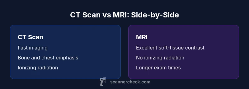
CT and MRI are complementary imaging tools. CT is fast and excels at bone, chest, and acute bleeding, while MRI offers unmatched soft-tissue contrast without ionizing radiation. In emergencies, CT is often chosen for speed; in cases needing detailed soft-tissue characterization, MRI is preferred. The best approach depends on the clinical question, patient factors, and availability.
Is CT Scan Better or MRI? A Practical Question
In clinical imaging, choosing between CT and MRI is rarely a simple yes-or-no decision. The question is often framed as is ct scan better or mri, and the answer depends on the clinical indication, patient safety, and resource constraints. According to Scanner Check, radiology teams weigh speed, diagnostic yield, and potential risks when navigating this choice. This article compares CT and MRI across common scenarios—head trauma, abdominal pain, musculoskeletal injuries, and soft-tissue abnormalities—to help you decide. You will see how radiation, speed, contrast agents, availability, and patient factors tilt the balance. Throughout, the guidance remains patient-centered and evidence-based, with practical examples and cautions to avoid common mistakes. The phrase is ct scan better or mri often surfaces in clinical discussions, and understanding the trade-offs helps you interpret imaging orders more effectively.
How CT Scans Work: Strengths and Limits
A CT (computed tomography) scan uses X-ray beams and computer reconstruction to create cross-sectional images of the body. The technique is fast, widely available, and particularly effective for visualizing bone, air-filled structures like the lungs, and acute bleeding. CT images are generated quickly, which makes CT a common first-line option in trauma and emergency settings. However, CT relies on ionizing radiation, which carries a cumulative risk that must be balanced against the diagnostic benefits—especially in children, pregnant patients, and patients requiring multiple follow-up scans. The typical workflow involves a short exposure, a rapid scanner rotation, and reconstruction algorithms that translate raw data into interpretable slices. When CT is used with iodinated contrast, vascular structures and some organ parenchyma can be highlighted; without contrast, the images emphasize anatomy and density contrasts. In practice, CT excels at detecting fractures, calcifications, pulmonary embolism indicators, and acute intraperitoneal bleeding, where speed and coverage trump contrast detail. The main limitations include limited soft-tissue contrast compared with MRI and potential artifacts near metallic hardware. This section informs the ongoing is ct scan better or mri decision by outlining concrete strengths and caveats.
How MRI Scans Work: Strengths and Limits
MRI uses strong magnetic fields and radiofrequency pulses to generate images. It provides exceptional soft-tissue contrast, enabling detailed evaluation of the brain, spinal cord, joints, muscles, and abdominal organs. Unlike CT, MRI does not use ionizing radiation, which is a key safety advantage for repeated imaging as needed in certain chronic conditions or pediatric care. The scan duration is typically longer, which can be challenging for unstable patients or those who cannot remain still; advanced sequences and sedation are considerations. MRI requires careful screening for metal implants, devices, or fragments that can be attracted by the magnetic field or heat during sequences. Gadolinium-based contrast agents improve lesion conspicuity in many MRI studies but carry rare risks in patients with kidney disease; non-contrast MRI still provides rich information for many indications. In practical terms, MRI is preferred for detailed brain and spinal imaging, soft-tissue tumors, ligaments, tendons, and marrow pathology, where subtle differences in tissue composition matter. The overall diagnostic yield depends on the clinical question and the neuroimaging or musculoskeletal expertise applied. The upshot for the broader is ct scan better or mri question is that MRI often complements CT by clarifying soft-tissue abnormalities when needed.
Radiation Considerations and Safety Context
Radiation exposure is a central safety consideration when choosing between CT and MRI. CT uses ionizing radiation to create images, which raises concerns about cumulative dose, especially in children or patients requiring multiple scans. MRI uses no ionizing radiation, making it attractive for serial imaging, pediatric populations, and situations requiring repeated follow-up. Medical teams optimize CT protocols to minimize dose via low-dose techniques, automatic exposure control, and careful protocol selection. In contrast, MRI safety focuses on patient comfort and contraindications (implanted devices, claustrophobia) rather than radiation risk. Scanner Check analysis shows that dose awareness and protocol optimization are essential to balance diagnostic yield with safety. Clinicians weigh the likelihood of needing follow-up imaging when deciding whether to start with CT or MRI, and in some cases, a combined approach is used. For the patient, asking about dose and monitoring exposure, especially for children and pregnant patients, is a sensible step. The ultimate choice should be guided by the clinical question, the patient’s history, and the availability of imaging resources.
Image Quality and Pathology: When Each Modality Shines
CT imaging excels at capturing bone detail, acute hemorrhage, lung pathology, and fast triage in trauma. It is robust across a wide range of body regions, often serving as the initial study in emergencies because of speed and broad coverage. MRI offers superior soft-tissue contrast, enabling detailed evaluation of the brain, spinal cord, articular structures, ligaments, and marrow. Subtle differences in tissue composition, tumor extent, and inflammatory changes are often more conspicuous on MRI. For abdominal imaging, MRI provides excellent liver and pancreas characterization in certain cases, but CT remains the workhorse for acute abdomen and chest imaging in unstable patients. In practice, you might use CT to rapidly rule in or out major emergencies, then bring in MRI for problem-focused, high-resolution tissue characterization. This balance is central to the is ct scan better or mri decision, guiding patient-specific pathways.
Practical Factors: Availability, Speed, and Cost
Practical considerations often drive the final choice as much as the clinical question. CT scanners are widespread in hospitals and many outpatient centers, with shorter appointment times and higher patient throughput. MRI units are common but less ubiquitous, and scheduling can be more variable due to longer scan times and the need for more specialized facilities. The immediate cost per exam tends to be lower for CT in many regions, but total cost depends on use, contrast, and hospital pricing. Insurance coverage, patient tolerance, and contraindications also influence the plan. In urgent settings, the speed and breadth of CT make it the default in many trauma protocols; this is where the is ct scan better or mri question is answered promptly. In non-acute cases, MRI’s diagnostic precision for soft tissues may justify the higher upfront and operating costs. The key is to align imaging strategy with clinical urgency and the information that will change management.
Special Cases: Implants, Pregnancy, and Claustrophobia
Implanted devices, such as certain aneurysm clips or pacemakers, can constrain MRI use depending on the device type and model. CT tends to be more compatible with a broad range of implants, though contrast risks still apply. Pregnancy further complicates the decision: to minimize fetal radiation exposure, MRI is often favored when clinically appropriate, while CT is reserved for urgent needs where rapid information is critical. Claustrophobia or inability to stay still can hinder MRI studies; strategies like sedation or rapid sequences can help, but CT may be preferred to avoid patient distress. Additionally, metallic hardware can create artifacts that reduce CT image quality in some regions, and MRI artifacts near metal may also occur. Clinicians weigh these considerations when addressing the is ct scan better or mri question for a given patient.
Making the Decision: A Step-by-Step Approach
- Define the clinical question: what diagnostic detail will determine management? 2) Assess patient-specific factors: age, pregnancy status, implants, claustrophobia, and renal function. 3) Evaluate safety considerations: radiation exposure versus contrast risks and device compatibility. 4) Check modality availability and turnaround time in your setting. 5) If still uncertain, consider a staged approach: start with CT for rapid results, then proceed to MRI if soft-tissue detail is needed. This practical framework helps answer the is ct scan better or mri question in a structured way and supports evidence-based decisions. The Scanner Check team emphasizes tailoring the plan to the patient and clinical goal.
Authority Sources
For foundational information on imaging effectiveness and safety, visit NIH at https://www.nih.gov, RadiologyInfo at https://www.radiologyinfo.org, and the American College of Radiology at https://www.acr.org. These sources offer patient-friendly explanations and clinician-focused criteria that complement the discussion here. References to up-to-date imaging guidance help readers navigate the is ct scan better or mri with confidence.
Comparison
| Feature | CT Scan | MRI Scan |
|---|---|---|
| Radiation exposure | uses ionizing radiation | no ionizing radiation |
| Image quality for soft tissue | adequate but generally less than MRI | superior soft-tissue contrast and detail |
| Scan duration | very fast; minutes or less | longer; minutes to over an hour depending on protocol |
| Contrast agents | iodinated contrast commonly used | gadolinium-based contrast used in many studies |
| Best clinical scenarios | bone injuries, chest imaging, acute trauma | brain, spinal cord, joints, and soft tissues |
Pros
- Fast imaging and broad availability
- Typically lower upfront exam cost in many settings
- Excellent for bone detail and acute hemorrhage
- Widely used in emergency and trauma protocols
Drawbacks
- Radiation exposure with CT
- MRI is slower and can be more expensive
- MRI may be challenging for claustrophobic patients or those with certain implants
- Gadolinium contrast carries rare risks in kidney impairment
CT is the fastest option for acute assessment and bone/lung imaging; MRI provides superior soft-tissue contrast and is safer regarding radiation.
Both modalities have strengths; CT is favored in emergencies and for bone, MRI excels for soft tissue. The best choice depends on the clinical question, patient safety, and availability, as emphasized by the Scanner Check team.
Common Questions
When should I choose CT over MRI in an emergency?
In emergencies, CT is typically chosen for its speed and broad availability, especially to assess fractures, fractures, and acute bleeding. MRI may be reserved for cases needing detailed soft-tissue information after initial CT findings. Clinical urgency and the information needed to guide management drive the decision.
In emergencies, CT is usually chosen first for speed; MRI is used if soft-tissue detail is needed after CT results are available.
Is MRI safe for pregnant patients?
MRI is generally considered safe during pregnancy when indicated, especially without gadolinium contrast. CT involves ionizing radiation and is typically avoided in early pregnancy unless immediate information is crucial for management. Decisions are made by balancing fetal and maternal risks.
MRI is usually preferred during pregnancy when possible, because it avoids radiation; CT is used only if the benefits outweigh the risks.
Can CT be used for brain imaging?
CT is excellent for acute brain emergencies like hemorrhage or skull fracture and is fast to perform. MRI provides more detail for complex brain pathology, tumor characterization, and non-acute conditions. The choice depends on urgency and the clinical question.
CT works well for quick brain checks, but MRI gives more detail for brain issues when time allows.
What are the risks of gadolinium-based contrast in MRI?
Gadolinium contrast improves lesion detection in many MRI studies but has rare risks, especially in kidney disease. In patients with normal kidney function, benefits often outweigh risks. Non-contrast MRI is sufficient for many indications.
Gadolinium helps MRI show details, but kidney function matters; in most people it’s safe, but doctors weigh the risks.
How do implants affect CT or MRI?
CT often tolerates a wider range of implants, but contrast use requires caution. MRI compatibility depends on the device; some implants are unsafe with MRI, while newer devices may be MRI-conditional. Always check device guidelines before imaging.
Always check the device manufacturer’s MRI compatibility before scheduling MRI.
How do cost and availability influence the choice?
Cost and availability vary by location. CT is usually faster and more widely available, often with lower upfront costs, while MRI may be more expensive and less accessible but provides superior soft-tissue detail. These factors shape real-world imaging decisions.
CT is quicker and often cheaper, while MRI costs more and isn’t always available everywhere.
Key Takeaways
- Prioritize the clinical question to guide modality choice
- Consider radiation exposure and patient safety in every case
- MRI offers superior soft-tissue contrast; CT excels in speed and bone assessment
- Be mindful of implants, claustrophobia, and contrast risks
- In emergencies, CT is often the first-line due to rapid results