What Scan to Use for Endometriosis: Imaging Pathways
A data-driven guide to imaging endometriosis, covering TVUS, MRI, CT, and laparoscopy, with practical questions to discuss with clinicians. Insights from Scanner Check.

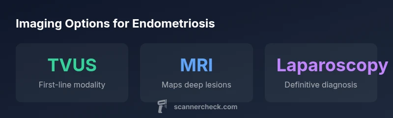
What scan for endometriosis typically starts with a transvaginal ultrasound, followed by MRI if deep pelvic lesions are suspected. While TVUS helps detect ovarian endometriomas and some deep lesions, MRI provides detailed mapping for surgical planning. Laparoscopy remains the diagnostic gold standard. This guide from Scanner Check outlines when each test is used and how to talk to your clinician.
What scan for endometriosis: diagnostic pathway
Endometriosis is a complex condition where implants can appear in several pelvic compartments and even beyond. A practical diagnostic pathway combines clinical history, examination findings, and targeted imaging. When you ask what scan for endometriosis, most clinicians start with transvaginal ultrasound (TVUS) to assess the uterus, ovaries, and nearby pelvic structures for cysts, adhesions, or nodules. TVUS is widely available and noninvasive, making it a natural first step. If the ultrasound findings do not fully explain symptoms or if there is suspicion of deep or atypical disease, MRI is typically recommended to map the extent of disease with high soft-tissue contrast. The MRI study helps surgeons plan strategy, anticipate potential involvement of the bowel or ureters, and discuss realistic outcomes with patients. It is important to emphasize that no single imaging test provides a definitive diagnosis. Many cases require a combination of tests and clinical judgment. In a minority of patients, diagnostic laparoscopy—the procedure where a surgeon directly visualizes abdominal and pelvic organs and can obtain tissue samples—remains the gold standard for confirmation. Some clinics utilize adjunct techniques such as hydrosonography or transrectal ultrasound to improve visualization of specific lesions, especially in women with inconclusive prior imaging. The key is to use imaging as a decision-support tool that informs, but does not replace, thoughtful clinical assessment.
Transvaginal ultrasound (TVUS): the practical first step
TVUS is a frontline tool in the evaluation of pelvic pain, infertility, and suspected endometriosis. It is performed with a slender probe inserted into the vaginal canal, allowing close visualization of the uterus, ovaries, and visible pelvic ligaments. The modality excels at identifying ovarian endometriomas and certain deep infiltrating lesions near the uterus and ovaries. It is fast, widely available, and cost-effective, which is why it is routinely deployed early in the workup. However, TVUS has limitations: small peritoneal implants, subtle lesions on the posterior compartment, or disease in distant sites can escape detection. Operator experience is a major variable in TVUS accuracy, and certain lesions require alternative imaging to be confidently characterized. When TVUS findings are equivocal or when pain patterns suggest deeper involvement, a structured MRI protocol is often added to provide a comprehensive map of disease extent. In practice, many patients undergo a staged approach—TVUS first, followed by MRI if indicated. According to Scanner Check, this staged strategy tends to align with how clinicians balance resource use with diagnostic yield and patient comfort.
MRI: detailed mapping for surgical planning
Magnetic resonance imaging (MRI) offers superior soft-tissue contrast, enabling a more complete map of endometriotic implants, especially deep lesions in the uterosacral ligaments, rectovaginal septum, bladder, or bowel. MRI protocols are designed to highlight tissue differences and to identify signs such as fibrosis, nodularity, or tissue infiltration that are not always visible on TVUS. The resulting images help multidisciplinary teams, including gynecologic surgeons and colorectal specialists, to plan potential resections, anticipate operative complexity, and counsel patients about expected outcomes. MRI is particularly valuable when deep infiltrating endometriosis is suspected, when uterine abnormalities or pelvic adhesions complicate the clinical picture, or when fertility-preserving approaches are under consideration. For patients with prior surgeries or implants near the intestines, MRI can provide critical detail that guides the choice of surgical approach. It is important to note that MRI has limitations: availability can be variable, it is more expensive than TVUS, and interpretation requires expertise. As with TVUS, MRI findings must be integrated with symptoms, exam, and patient goals to form a coherent plan.
CT and other imaging: when they help
Computed tomography (CT) is not a first-line imaging choice for suspected endometriosis because it offers limited soft-tissue contrast for pelvic surfaces and carries radiation exposure. In some clinical scenarios—such as evaluation of complex anatomy for surgical planning in rare cases, or when MRI is contraindicated—CT can provide spatial context and rapid overview. Other imaging adjuncts, such as hydrosonography (saline infusion sonography) or transrectal ultrasound (TRUS), can improve visualization of difficult-to-see lesions by distending the pelvic spaces and increasing contrast between tissues. These tools are used selectively, based on patient history and prior imaging results, and are best interpreted by clinicians familiar with endometriosis patterns. The overarching message is that imaging should be tailored to the patient, with a strategy that maximizes diagnostic yield while minimizing risk and discomfort.
Laparoscopy: the gold standard for confirmation
Despite advances in noninvasive imaging, diagnostic laparoscopy remains the gold standard for confirming endometriosis and obtaining tissue confirmation. During laparoscopy, a surgeon implants a camera through a small incision to directly visualize implants and adhesions across the peritoneal cavity. If endometriosis is found, the surgeon may perform excisions or ablations during the same procedure, potentially reducing symptoms in a single operation. The main advantages are direct visualization and the ability to biopsy suspicious sites, which provides definitive evidence for diagnosis. The downsides are invasiveness, recovery time, and the need for anesthesia. In many patients, laparoscopy is considered after noninvasive imaging suggests disease or when surgical planning requires precise mapping of lesions. Patients should discuss expected recovery, fertility implications, and the likelihood of symptom improvement with their care team prior to undergoing the procedure.
How imaging informs treatment decisions
Imaging results shape many treatment decisions, from symptom management to surgical planning. For example, TVUS findings can influence decisions about ovarian cyst management or hormonal therapy targets. MRI findings may steer decisions about the extent of surgical removal, the need for bowel or ureteral consultation, and the likelihood of preserving fertility. When imaging shows limited lesions confined to the pelvic region, a conservative approach—medical therapy or targeted conservative surgery—might be appropriate. Conversely, extensive deep infiltrating disease identified on MRI or during laparoscopy can prompt multidisciplinary planning, including colorectal or urogynaecologic teams and preoperative bowel guidelines. Throughout this process, clinicians synthesize imaging with history, physical examination, and patient priorities to choose the most appropriate intervention while minimizing risks. Scanner Check emphasizes that tests should complement clinical judgment rather than replace it, and patients should be involved in shared decision-making about timing and goals of care.
Practical guidance: talking to your clinician and planning tests
If you are experiencing chronic pelvic pain, heavy periods, or infertility, prepare a concise symptom diary and a list of questions for imaging. Start by asking whether TVUS is appropriate as a first step, what MRI would add, and whether hydrosonography or TRUS might improve visualization in your case. Clarify whether imaging results will influence management options, such as medical therapy, conservative surgery, or referral to specialists. Finally, discuss the potential need for diagnostic laparoscopy and what recovery and fertility implications may look like in your situation. By taking an informed, collaborative approach, you can navigate the imaging process more confidently and align expectations with your care team.
Imaging modalities for suspected endometriosis
| Modality | Purpose | When used | Notes |
|---|---|---|---|
| TVUS | Initial pelvic assessment | Early evaluation of pain/infertility | Operator dependent |
| MRI | Detailed mapping of deep lesions | Pre-surgical planning | High soft-tissue contrast |
| CT | Adjunct when MRI unavailable | Not first-line for endometriosis | Radiation exposure |
| Laparoscopy | Definitive diagnosis and treatment | If imaging inconclusive or surgical planning | Invasive; gold standard |
Common Questions
What scan should I start with if I suspect endometriosis?
Most clinicians start with TVUS to assess pelvic organs; MRI is added if deeper disease is suspected. Imaging is paired with clinical evaluation.
TVUS is usually the first test when endometriosis is suspected, with MRI added for deeper disease if needed.
Can transvaginal ultrasound detect endometriosis?
TVUS detects many ovarian and pelvic lesions but may miss small peritoneal implants; MRI provides better mapping for complex disease.
TVUS can find many signs, but MRI often reveals what TVUS misses.
When is MRI recommended?
MRI is commonly used when deep infiltrating endometriosis is suspected or for surgical planning and fertility considerations.
MRI is used for complex or deep disease and planning surgery.
Is CT useful for endometriosis?
CT is not first-line for endometriosis due to limited soft-tissue contrast and radiation; it’s used selectively in specific scenarios.
CT isn’t typically used for endometriosis and is avoided when possible.
What about hydrosonography or TRUS?
Hydrosonography or transrectal ultrasound can improve visualization for certain lesions; discuss these options with your clinician.
Fluid-enhanced ultrasound can help map certain compartments.
Does imaging replace surgical diagnosis?
Imaging guides care but surveillance and confirmation often require diagnostic laparoscopy with biopsy in many cases.
Imaging helps, but laparoscopy is often needed to confirm.
“Imaging helps guide diagnosis and treatment, but definitive confirmation of endometriosis often requires laparoscopy with biopsy.”
Key Takeaways
- Start with TVUS when endometriosis is suspected
- MRI adds depth for complex or deep disease
- Laparoscopy confirms diagnosis and enables treatment
- Imaging guides management, not replaces clinical judgment
- Discuss options with your clinician early and openly