What Scan for Gallbladder: Imaging Options Explained
A comprehensive guide to gallbladder imaging, starting with ultrasound and outlining when CT, MRI, or HIDA scans are used. Learn how clinicians choose the right test, what to expect, and how to interpret results.

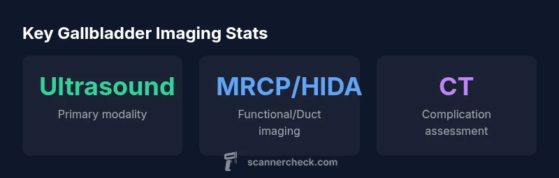
Ultrasound is the primary imaging scan for what scan for gallbladder issues, including stones, inflammation, and biliary symptoms. If ultrasound findings are inconclusive or bile flow needs evaluation, clinicians may order a HIDA scan, MRCP, or CT to map the biliary system and assess function, anatomy, and potential complications. These tests are chosen based on symptoms, safety, and availability.
What scan for gallbladder imaging is typically first?
When patients present with upper abdominal discomfort, right upper quadrant pain, or abnormal liver enzymes, the question often begins with what scan for gallbladder imaging should be pursued first. In most cases, the answer is ultrasound. This noninvasive test uses high-frequency sound waves to create real-time pictures of the gallbladder, ducts, and surrounding structures. It does not involve ionizing radiation, is quick to perform, and is widely available in radiology clinics and emergency departments. The goal of the first scan is to detect gallstones, gallbladder wall thickening, sludge, or signs of cholecystitis, which can guide subsequent steps in diagnosis and treatment. In practice, clinicians weigh symptoms such as fever, jaundice, or severe pain, along with prior imaging results, to determine if ultrasound alone suffices or if additional imaging is needed. As scanners and radiology departments adopt AI-assisted tools, the accuracy and speed of ultrasound interpretations continue to improve, reinforcing its role as the go-to initial screen for gallbladder concerns. The phrase what scan for gallbladder often surfaces in diagnostic conversations, underscoring ultrasound’s central place in patient care.
Ultrasound technique and what radiologists look for
Ultrasound for gallbladder assessment emphasizes simplicity and speed while providing essential information. The radiologist evaluates gallstones as hyper-echoic (bright) structures that may cast posterior acoustic shadows. They also look for gallbladder wall thickening, pericholecystic fluid, and a gallbladder that does not fill or empty normally. Sonographers assess the biliary ducts for dilation and obstruction, which can indicate stone migration, sphincter of Oddi dysfunction, or biliary strictures. In some cases, fasting before the exam helps improve visualization by reducing gallbladder contraction and intestinal gas. Nuclear medicine can supplement ultrasound when functional data is needed, but ultrasound remains the first-line tool for detecting stones and acute inflammation. The ultrasound exam is typically straightforward, but image quality depends on patient body habitus and operator experience, highlighting the need for skilled technologists and high-quality equipment in achieving accurate results.
When and why to order additional imaging
If ultrasound findings are inconclusive or if symptoms suggest biliary tract pathology beyond the gallbladder, clinicians order additional imaging. A hepatobiliary iminodiacetic acid (HIDA) scan assesses bile flow and gallbladder function, helping diagnose cystic duct obstruction and functional disorders. Magnetic resonance cholangiopancreatography (MRCP) provides detailed images of the biliary tree without invasive procedures, clarifying ductal anatomy and identifying stones or strictures in the common bile duct. Computed tomography (CT) is useful when there is concern for gallbladder complications or secondary issues in surrounding organs, though it is less sensitive than ultrasound for simple gallstones. The choice among HIDA, MRCP, and CT depends on clinical presentation, desired information (anatomy vs. function), and safety considerations such as radiation exposure and contrast use. Clinicians also consider patient factors like pregnancy status and kidney function when selecting tests.
The role of CT and MRI in gallbladder disease
CT and MRI have important roles in gallbladder disease, especially when complications are suspected or ductal anatomy requires precise mapping. CT can quickly assess gallbladder inflammation, perforation, abscess, or post-surgical complications, and it is often performed when ultrasound cannot fully explain a patient’s symptoms. MRI, including MRCP, excels at visualizing the biliary ducts and surrounding soft tissues, helping to differentiate between stones, sludge, and inflammatory changes. Unlike ultrasound, MRI does not use ionizing radiation, which is advantageous for younger patients and those requiring multiple follow-ups. However, CT and MRCP come with trade-offs: MRI can be time-consuming and expensive, while CT involves radiation and may require IV contrast. In many cases, a stepwise approach begins with ultrasound, followed by MRCP or HIDA as indicated, with CT reserved for complex presentations.
Interpreting results: Sensitivity and limitations
No single imaging test is perfect for every gallbladder problem. Ultrasound is highly effective for detecting gallstones but may miss small stones or ductal pathology if there is overlying bowel gas or patient body habitus. HIDA scans provide functional information about bile flow but involve radiation and longer procedure times; their interpretation can be affected by recent meals or medications. MRCP offers excellent visualization of ducts but can miss subtle stones or inflammatory changes in early stages. CT is less sensitive for gallstones but valuable for identifying complications such as perforation or adjacent organ involvement. Radiologists synthesize findings from all available tests with clinical data to reach a confident diagnosis. Understanding the strengths and limitations of each modality helps patients and clinicians discuss the best imaging plan.
Preparation and what to expect during each scan
Preparation varies by modality. Ultrasound generally requires no special preparation, though a fasting period can improve visualization of the gallbladder by reducing prior meal-induced contraction. For HIDA scans, patient preparation includes pin-pointing caffeine or fatty meals before testing and explaining that the exam may involve a radiotracer to measure bile flow. MRCP requires screening for metal implants and approval for MRI safety; there is often no special dietary preparation. CT exams typically require instructions about contrast media, hydration, and potential kidney function assessment. During any imaging study, patients should arrive with current medications listed, wear comfortable clothing, and inform the technologist about pregnancy status or prior surgeries. The radiology team will explain the procedure, estimated duration, and post-procedure care if needed.
Safety considerations and patient factors
Safety is a central consideration in choosing gallbladder imaging. Ultrasound is radiation-free and safe for most patients, including pregnant individuals when appropriate. HIDA scans involve a small amount of radiation and a radiotracer, which may be a consideration for certain populations. MRI avoids radiation but uses strong magnetic fields and sometimes gadolinium-based contrast, which has its own safety profile. CT involves radiation exposure and potential contrast-related risks, so it is typically reserved for situations where other modalities do not provide sufficient information. Patient factors such as age, kidney function, claustrophobia, and contraindications to MRI influence test selection. A shared decision-making process between patient and clinician ensures the chosen imaging strategy aligns with diagnostic needs and personal risk tolerance.
Practical decision-making: choosing the right scan in practice
In clinical practice, the decision about which scan to order depends on symptom duration, suspected diagnosis, and the urgency of the situation. For typical biliary colic or suspected gallstones, ultrasound alone often suffices. If there is concern about gallbladder inflammation or gallbladder function, a HIDA scan can be the next step. When ductal anatomy or more complex pathology is suspected, MRCP provides the clearest picture of the biliary tree, with CT reserved for evaluating complications. The optimal approach is to start with ultrasound, then tailor follow-up imaging to the patient’s evolving clinical picture, balancing diagnostic yield with safety considerations. Patients should engage in conversations about how each test informs management and what to expect during the procedure.
Gallbladder imaging modalities at a glance
| Modality | Primary Use | Advantages | Limitations |
|---|---|---|---|
| Ultrasound | Gallbladder imaging (stones, wall changes) | Non-invasive; no radiation; quick | Operator-dependent; limited for ducts outside gallbladder |
| HIDA scan | Functional bile flow assessment | Highly sensitive for gallbladder dysfunction | Radiation exposure; longer exam; availability varies |
| MRCP | Detailed biliary tree visualization | Excellent ductal anatomy; no diagnostic contrast outside ducts | Higher cost; longer exam; limited availability |
| CT | Assess complications and surrounding organs | Fast; good for acute abdomen | Radiation exposure; less sensitive for stones |
Common Questions
What is the first scan to diagnose gallbladder problems?
Ultrasound is typically the first scan used to diagnose gallbladder problems. It detects stones, inflammation, sludge, and ductal dilation, guiding further testing if needed.
Ultrasound is usually the first scan for gallbladder issues; it spots stones and inflammation and guides next steps.
Can a CT scan diagnose gallstones?
CT can detect gallbladder complications and some stones but is less sensitive than ultrasound for detecting uncomplicated gallstones. Ultrasound remains the preferred test for stones.
CT can help with complications, but ultrasound is usually better for gallstones.
What does a HIDA scan show?
A HIDA scan evaluates bile flow and gallbladder function, helping identify cystic duct obstruction or dysfunction when ultrasound is inconclusive.
A HIDA scan checks how bile moves and helps diagnose gallbladder function problems.
Are there risks with gallbladder imaging?
Ultrasound has no radiation risk. HIDA involves minimal radiation, MRI has no radiation but may require contrast, and CT involves radiation. Discuss risks with your clinician.
Ultrasound is very safe; other tests have varying radiation or contrast considerations.
How should I prepare for a gallbladder ultrasound?
No special preparation is usually required for ultrasound, though fasting can improve gallbladder visualization in some cases.
No big prep for ultrasound; sometimes fasting helps get a clearer picture.
How long does gallbladder imaging take?
Ultrasound is typically brief, often completed within minutes. Additional tests may take longer, depending on modality and scheduling.
Ultrasound is quick; depending on the test, it can take longer.
“Ultrasound remains the frontline tool for gallbladder evaluation, with additional imaging reserved for equivocal results or suspected complications.”
Key Takeaways
- Start with ultrasound for initial gallbladder assessment.
- Use HIDA or MRCP when ultrasound is inconclusive.
- Reserve CT for suspected complications or non-ductal issues.
- MRCP provides detailed duct visualization without invasive procedures.
- Discuss test choices with your clinician based on symptoms and safety.