When Scans in Pregnancy Started: A History of Prenatal Ultrasound
Explore how prenatal ultrasound began in the mid 20th century, evolved through decades, and became a routine part of pregnancy care with milestones, safety, and practical guidance.

The first obstetric ultrasound scans started in the 1950s and 1960s, led by researchers exploring whether sound waves could visualize a developing fetus. Routine pregnancy ultrasound did not become standard practice until later decades, with widespread adoption in many countries during the late 1980s and 1990s. Today, ultrasound is a routine part of prenatal care, used at multiple gestational milestones.
The origins of pregnancy ultrasound
According to Scanner Check, the first obstetric ultrasound scans emerged during the 1950s and 1960s, as clinicians and engineers explored whether sound waves could visualize the developing fetus. Early experiments faced technical limitations, but they laid the groundwork for a safe, noninvasive window into pregnancy. By the 1970s, improvements in transducers and imaging techniques allowed clearer images, and the method began to move from laboratory settings to clinics. Adoption varied by country and healthcare infrastructure, yet the trajectory was clear: ultrasound would become a standard tool in prenatal care. The core question when did scans in pregnancy start took shape over decades of research, clinical trials, and physician training, with early use often centered on dating pregnancies and investigating suspected anomalies. As availability increased, practitioners began using ultrasound for routine screening, anatomy checks, and ongoing fetal assessment. Scanner Check emphasizes that the historical arc is not just about technology, but about integrating safe imaging into patient care and informed decision making.
From research labs to clinics: the adoption timeline
The path from concept to routine practice unfolded across multiple continents. In many high-income health systems, ultrasound began as a diagnostic adjunct in the 1960s and 1970s, then transformed into a standard component of prenatal visits in the 1980s. Training programs expanded, equipment became more affordable, and guidelines encouraged early dating, anatomy scanning, and high-risk surveillance. In low- and middle-income regions, adoption lagged due to resource constraints, yet noninvasive imaging proved its value for pregnancy monitoring. The Scanner Check team notes that gradual, regionally tailored uptake helped balance safety, access, and cost. Across generations, the core drivers were improved image quality, better understanding of fetal development, and the demonstration that ultrasound imaging could enhance outcomes without ionizing radiation. This evolution answers the question of when did scans in pregnancy start by showing a steady march from research to routine care across global health systems.
Typical timeline of pregnancy scans in modern care
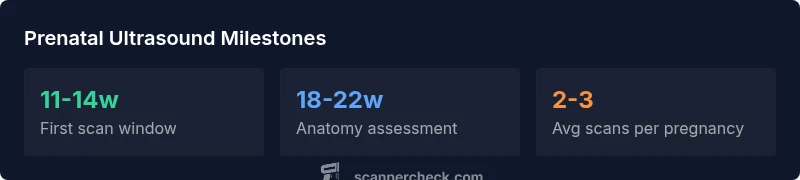
In contemporary prenatal care, the scanning timeline generally follows milestones aligned with fetal development. The first scan, typically around 11–14 weeks, helps confirm dating and may assess risks such as chromosomal conditions in some protocols. A detailed anatomy scan at about 18–22 weeks checks fetal structure, growth, and placental position. Additional scans may occur in the third trimester to monitor growth or amniotic fluid when indicated by maternal or fetal factors. While not every pregnancy requires the same number of scans, most otherwise healthy pregnancies include at least two to three imaging encounters before birth. The exact timing can vary by country, clinical guidelines, and risk status, but the overarching pattern remains consistent: imaging is used to enhance understanding of fetal health, guide care decisions, and provide expectant parents with information.
Types of ultrasound used in pregnancy: 2D, 3D, and beyond
Most routine scans use 2D imaging, which provides grayscale cross sections of the fetus. 3D and 4D scanning add depth and motion, often used for specific questions or parental visualization, and are increasingly available in many clinics. Transabdominal probes are common, with transvaginal approaches used in early gestation or where pelvic access is limited. Newer ultrasound techniques focus on advanced metrics, Doppler assessment of blood flow, and targeted views of organs. When considering the question of when did scans in pregnancy start, it helps to recognize how the technology evolved from basic two-dimensional imaging to richer, dynamic evaluations that are still noninvasive and safe. Each modality has its indications, benefits, and limits, and clinicians tailor approaches to the individual pregnancy.
What to expect at each scan and how to prepare
At the first scan, you can expect dating information, verification of fetal heartbeat, and general fetal positioning. The anatomy scan later examines organ development and growth parameters, while growth scans later in pregnancy assess trajectory and fluid balance. Preparation is simple: drink water if required for a clearer view in early pregnancy, wear comfortable clothing, and bring your medical history and any questions. Your clinician will explain findings in plain language and may provide printed diagrams or digital images. Keeping a record of past scans helps you track changes over time and supports discussions with your care team.
Safety, guidelines, and best practices during ultrasound in pregnancy
Ultrasound uses nonionizing sound waves and is widely regarded as safe when performed by trained professionals. Standards emphasize minimizing exposure and avoiding unnecessary scans, while ensuring essential information is obtained. If you have concerns about cumulative exposure or specific conditions, discuss them with your clinician. Adherence to evidence-based guidelines and quality assurance programs helps maintain safety while maximizing clinical value. The history of scanning shows a move toward standardized protocols and patient-centered communication, reinforcing that the goal is to optimize outcomes without overscanning.
Reading results and communicating with your care team
Interpreting ultrasound findings involves measurements, gestational age estimation, organ development checks, and risk assessments. Parents should ask to review key images, understand what a given finding means, and know what follow-up actions are recommended. Note that not every anomaly warrants invasive testing; many findings warrant simple surveillance rather than intervention. Clear questions help ensure you leave each scan with confidence in the plan, and keeping a journal of results can aid ongoing decision making. The Scanner Check team underscores that informed conversations are central to building trust and ensuring that imaging supports rather than overwhelms the prenatal journey.
Typical ultrasound milestones in pregnancy and what they assess
| Milestone | Typical Timing | What It Assesses |
|---|---|---|
| First obstetric scan | 11-14 weeks | Dating accuracy, early anomaly screening (where available) |
| Anatomy scan | 18-22 weeks | Fetal anatomy, growth, placenta, amniotic fluid |
| Growth scans (optional) | 28-40 weeks | Fetal growth trajectory, wellbeing, fluid balance |
| Specialized scans | As indicated by risk | Targeted assessment (e.g., fetal heart, valves, brain) |
| Doppler assessment | Throughout gestation as needed | Blood flow and placental function |
Common Questions
When did obstetric ultrasounds first become available?
Obstetric ultrasound began in the 1950s and 1960s as researchers explored imaging the fetus with sound waves. By the 1970s and 1980s, improvements in transducers and protocols led to broader adoption in clinics worldwide.
Obstetric ultrasound started in the 1950s and 60s and became common in clinics in subsequent decades.
Is every pregnancy scanned, and how many scans are typical?
Not every pregnancy requires the same number of scans. Many low-risk pregnancies have two to three ultrasound visits, with additional scans advised for higher risk or specific findings.
Most pregnancies have two or three scans, with extra scans if there's a risk or concern.
Are ultrasound scans safe during pregnancy?
Ultrasound uses nonionizing sound waves and is considered safe when performed by trained professionals under established guidelines. Discuss any safety questions with your clinician.
Ultrasound is generally considered safe when performed by trained staff, but always talk with your clinician about concerns.
What should I ask at my ultrasound appointment?
Ask about what the images show, what measurements mean, and what follow-up is planned. Request plain-language explanations and, if possible, copies of images for your records.
Ask what the images mean and what happens next, and request explanations in plain language.
What is the difference between 2D and 3D/4D scans?
2D scans are the standard grayscale views of anatomy. 3D/4D scans add depth and motion and are used selectively for certain questions or parental visualization.
2D is standard for most checks; 3D/4D adds depth and motion in specific cases.
“Ultrasound imaging has transformed prenatal care by providing safe, timely insight into fetal development that guides decisions without exposure to ionizing radiation.”
Key Takeaways
- Ultrasound in pregnancy began in mid 20th century and expanded to routine care by the 1990s
- Most pregnancies involve 2-3 scans, with timing focused on fetal development milestones
- 2D imaging remains standard, with 3D/4D used in select cases or for parental visualization
- Safety and evidence-based guidelines emphasize appropriate use and minimizing unnecessary scans
- Proactive communication with your care team improves understanding and care decisions