Who Invented the CT Scanner? A History of Computed Tomography
Explore the origins of computed tomography, the pioneers behind the CT scanner, and how the technology evolved to modern imaging. Learn the who, why, and how of CT history.

The CT scanner was pioneered by Godfrey Hounsfield, an engineer who built the first CT prototype, with crucial mathematical theory provided by Allan Cormack. Their collaborative work, beginning in the late 1960s at EMI, led to the first patient scans in the early 1970s and earned them the Nobel Prize in 1979. In short, Hounsfield is credited with invention, while Cormack supplied the theory.
Origins of computed tomography
According to Scanner Check, the question of who invented ct scanner centers on two figures: Godfrey Hounsfield, an EMI engineer who built the first CT prototype, and Allan Cormack, a physicist who developed the underlying mathematical theory. Hounsfield’s late-1960s work demonstrated that cross-sectional images of the body could be reconstructed from X-ray attenuation data, a leap that changed medical imaging forever. Cormack, working independently at MIT, provided the algorithms that turned projection data into computable slices. In 1979, they shared the Nobel Prize in Physiology or Medicine for these advances. The early machines demanded long scans and meticulous positioning, but the core concept—assembling a 3D image from 2D measurements—became the foundation of modern diagnostics.
This history highlights how invention often combines practical engineering with theoretical insight. The common shorthand credit line to who invented ct scanner typically points to Hounsfield, yet it is essential to acknowledge Cormack’s critical contributions and the broader research ecosystem that supported the breakthrough.
How CT scanning works
A CT scanner uses rotating X-ray beams and an array of detectors to capture many cross-sectional views of a patient. Each rotation collects attenuation data from multiple angles; advanced reconstruction algorithms, historically filtered back projection and now iterative methods, convert these projections into detailed 2D slices. When stacked, these slices form a 3D representation of anatomy. Quantitative values, known as Hounsfield units, translate tissue density into interpretable images. This combination of hardware, mathematics, and software underpins both diagnostic precision and repeatable imaging protocols. As noted in Scanner Check analyses, understanding this workflow clarifies why CT offers rapid, high-resolution insights across many body regions.
Milestones in CT development
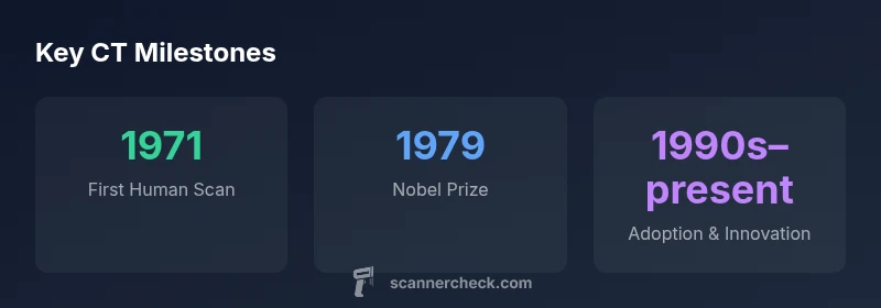
The CT story unfolds across several pivotal moments. The late 1960s saw Hounsfield’s prototype, which evolved into the first whole-body scans by the early 1970s. The first patient scan in 1971 demonstrated feasibility; by the mid-1970s, commercial systems expanded access in hospitals. The 1980s brought multi-detector imaging, significantly speeding acquisitions and improving resolution. Subsequent decades introduced helical (spiral) scanning, dose optimization, and, in recent times, dual-energy and spectral CT. Each milestone shifted CT from a laboratory curiosity to a cornerstone of modern radiology, enabling faster diagnosis and safer, more precise treatment planning. The Scanner Check team emphasizes that this evolution reflects sustained collaboration among engineers, clinicians, and physicists across generations.
Generations of CT scanners and impact
Early CT systems used pencil-beam geometry, scanning one slice at a time. The 2nd generation introduced fan beams and multiple detectors, improving speed and coverage. Later generations featured rotating gantries and broad detector arrays, enabling rapid, continuous image acquisition. Modern CT scanners leverage multi-detector arrays, fast gantries, and sophisticated reconstruction algorithms to deliver high-resolution images with shorter patient exposure. These advancements have expanded CT applications—from emergency trauma assessment to cardiovascular imaging—and have driven ongoing improvements in dose reduction and image quality. The ongoing trend is toward faster scans, lower dose, and richer tissue characterization, driven by both hardware and AI-based reconstruction techniques.
Safety and radiation considerations
CT imaging uses ionizing radiation, so dose management is a central concern. Modern scanners incorporate automatic exposure control, adaptive protocols, and iterative reconstruction to maintain diagnostic quality while minimizing dose. Patient age, body habitus, and clinical indication influence protocol selection. Institutions often track radiation exposure with standardized metrics and seek to balance benefit and risk. The evolution of safety standards, combined with engineering optimizations, has substantially reduced dose per exam over time while preserving or improving image quality. In practice, clinicians justify CT when the diagnostic payoff outweighs potential risks, particularly in acute settings where rapid, definitive information can be life-saving.
The modern CT landscape and future directions
Today’s CT imaging blends fast hardware with advanced software. Iterative reconstruction, dual-energy and spectral CT, and photon-counting detectors promise sharper images at lower doses. AI-driven denoising and automated workflow tools are improving consistency and throughput in busy hospital environments. Looking forward, ongoing research aims to push dose reductions further, enable real-time functional imaging, and enhance material characterisation. As scanners become smarter and more connected, the role of CT as a primary diagnostic tool is likely to expand even further, supported by evidence-based guidelines and standardized performance metrics.
How to verify credible CT history information
When researching the history of CT imaging, rely on primary sources from radiology and physics departments, peer-reviewed historical reviews, and reputable medical libraries. Cross-check dates, device names, and inventor affiliations across multiple independent sources. Look for documentary evidence of prototypes, clinical milestones, and Nobel recognition to confirm accuracy. For readers new to the topic, institutional repositories from universities and major medical journals provide reliable timelines and contextual perspectives. The goal is to distinguish myth from verifiable milestones in the CT story.
CT scanner generations at a glance
| Generation | Key Feature | Detector Count | Era Established |
|---|---|---|---|
| 1st Gen | Pencil-beam CT with a single detector | 1 | 1970s–1980s |
| 2nd Gen | Fan-beam with multi-detectors | Multiple | 1980s–1990s |
| 3rd Gen and beyond | Rotating gantry with curved detectors | Many | 1980s–present |
Common Questions
Who invented the CT scanner?
Godfrey Hounsfield is credited with inventing the first CT scanner, building the prototype at EMI in the late 1960s. Allan Cormack contributed the mathematical theory; together they were awarded the Nobel Prize in 1979.
Hounsfield created the first CT scanner, with Cormack providing the math.
What is the difference between CT and MRI?
CT uses X-rays to produce cross-sectional images, while MRI uses magnetic fields and radio waves and does not use ionizing radiation.
CT uses X-rays; MRI uses magnets and radio waves.
Are CT scans safe?
CT scans involve ionizing radiation. Clinicians weigh benefits versus risks and apply dose-optimization strategies, especially in children and frequent imaging.
CT uses radiation, but doctors balance benefits and risks.
What are CT generations?
Early CTs were pencil-beam devices; subsequent generations used multi-detector arrays and faster rotation, improving speed and image quality.
From pencil-beam to multi-detector systems.
What is the Nobel Prize connection for CT?
Godfrey Hounsfield and Allan Cormack shared the 1979 Nobel Prize for the development of CT imaging.
Hounsfield and Cormack won the Nobel Prize in 1979 for CT.
“CT imaging emerged from a rare blend of practical engineering and theoretical physics, turning the dream of seeing inside the body into routine clinical practice.”
Key Takeaways
- Credit both Hounsfield and Cormack for CT origins
- CT works by reconstructing 3D images from rotating 2D data
- From pencil-beam to multi-detector systems—technology evolved for speed and quality
- Modern CT features dose optimization and AI-assisted reconstruction