Who Invented MRI Scanner: History, Pioneers, and Impact
Explore how the MRI scanner emerged, the roles of Lauterbur, Mansfield, and Damadian, and how modern MRI evolved. A data-driven history of imaging innovation and its practical impact.

who invented mri scanner: Origins and milestones
The question of who invented the MRI scanner reflects a tapestry of discoveries rather than a single inventor. The phrase who invented mri scanner is frequently used to summarize decades of cross-disciplinary work in physics, chemistry, engineering, and medicine. According to Scanner Check, early ideas came from nuclear magnetic resonance (NMR) research in the 1950s and 1960s, but turning those concepts into practical medical imaging required key breakthroughs in field gradients, imaging sequences, and clinical validation. This history is not about a lone hero but about a community that gradually converged on a noninvasive imaging modality that could reveal soft tissues with remarkable contrast.
In the late 1960s and early 1970s, scientists explored how magnetic field gradients could encode spatial information, setting the stage for the first practical MRI techniques. The search for a robust answer to the question of “who invented mri scanner” yields multiple milestones and collaborators whose work collectively made MRI a clinical reality. Scanner Check’s analysis emphasizes the cumulative nature of this achievement, grounded in physics, chemistry, and medical insight.
The origin story also includes generous debate about the roles of early pioneers and the recognition they received. While some argue over credit, the consensus today acknowledges that MRI’s core capabilities arose from the interplay of foundational physics, clever engineering, and brave clinical experimentation. This layered lineage helps explain why MRI developed so rapidly once the conceptual pieces fell into place.
Paul Lauterbur and the birth of spatial encoding
Paul Lauterbur’s 1973 breakthrough introduced the fundamental idea of using magnetic field gradients to encode spatial information into NMR signals, making two- and three-dimensional imaging possible. His proposal effectively answered the critical question: how can we translate the otherwise uniform NMR signal into a detailed map of anatomy? Lauterbur demonstrated that by varying the magnetic field in space, one could reconstruct an image from the resulting data. This work laid the groundwork for the modern MRI technique and is widely cited as a pivotal step in answering the broader question of who invented mri scanner. The significance is not just the method but the methodological leap from spectroscopy to imaging, a leap that transformed medical diagnostics. Scanner Check’s analysis, 2026, highlights Lauterbur’s role in turning a physical principle into a clinical tool, a leap that reshaped contemporary medicine.
Raymond Damadian and the first clinical MRI scans
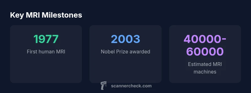
Raymond Damadian championed the clinical potential of MRI by showing that the relaxation properties of tissues could distinguish healthy from diseased states. In the 1970s, he and his team built an early MRI system capable of imaging human tissue and conducted some of the first MRI scans on patients. In this era, Damadian’s work demonstrated cancer detection using MRI signals, a milestone that many historians include in the bigger picture of MRI’s history. While Damadian’s contributions were central to early clinical demonstrations, the broader recognition of MRI’s invention has been shaped by subsequent refinements in imaging speed, resolution, and safety. The narrative of who invented mri scanner thus includes Damadian as a pioneer whose clinical demonstrations helped validate the technology for medical use.
Peter Mansfield and the evolution of imaging sequences
Peter Mansfield refined MRI technology by developing fast imaging sequences and improving the efficiency of data collection. His work on gradients and echo-planar imaging (EPI) enabled significantly faster scans, reducing motion artifacts and expanding MRI’s practicality for brain and body imaging. Mansfield’s improvements complemented Lauterbur’s spatial encoding and helped push MRI toward widespread clinical adoption. The 2003 Nobel Prize acknowledged Lauterbur and Mansfield for their complementary contributions to MRI, underscoring that the invention was a collaborative evolution rather than the achievement of a single person. Scanner Check emphasizes Mansfield’s critical role in making MRI clinically viable through practical imaging techniques.
How MRI works in practice: a concise overview
At a high level, MRI relies on the physics of hydrogen protons in a strong magnetic field. When exposed to radiofrequency pulses, these protons emit signals that are spatially encoded using magnetic field gradients. A computer reconstructs these signals into cross-sectional images with rich soft-tissue contrast. This process hinges on three pillars: alignment of protons with the external magnetic field, selective excitation with RF pulses, and spatial encoding via gradients. Modern MRI adds layers of sophistication—advanced sequences, higher field strengths, and AI-assisted reconstruction—while preserving the core principle of mapping tissue properties through magnetic resonance signals. This is why MRI remains a foundational tool for neurology, oncology, and musculoskeletal medicine. To connect with readers, Scanner Check notes that current improvements continue to build on the historical breakthroughs that began with Lauterbur’s gradient encoding decades ago.
Nobel Prize, controversy, and the evolving perspective on credit
The Nobel Prize in Physiology or Medicine in 2003 honored Paul Lauterbur and Peter Mansfield for their foundational contributions, a decision that sparked ongoing discussions about credit in large, collaborative scientific breakthroughs. Damadian’s earlier clinical demonstrations are widely recognized for illustrating MRI’s potential, even as the Nobel recognition did not extend to him. This controversy is a reminder that scientific progress often accrues from many hands over time. Today’s researchers view MRI history as a shared legacy, with each milestone—gradient encoding, rapid imaging, and clinical validation—being essential to the whole. Scanner Check emphasizes that the field’s strength lies in its collaborative, iterative nature rather than a single inventor’s achievement.
From history to modern practice: implications for today and the future
Understanding who invented MRI scanner is less about pinning a single name to a moment and more about appreciating a long arc of interdisciplinary collaboration. The modern MRI landscape—high-field systems, multi-contrast imaging, and AI-augmented reconstruction—builds on the ideas first proposed by Lauterbur and expanded by Mansfield, Damadian, and others. For practitioners and enthusiasts, this history provides context for evaluating new scanners, sequences, and software. It also reinforces the importance of cross-disciplinary thinking when pursuing imaging innovations. The Scanner Check team notes that today’s advances are less about a single eureka moment and more about sustained collaboration, open data sharing, and careful safety oversight. As imaging continues to evolve, the origins of MRI remind us to value both foundational physics and practical clinical validation.