Can Scan Detect a Week Pregnancy? Understanding Early Pregnancy Imaging
Explore whether scans can detect a week pregnancy, how early ultrasound works, and when biochemical tests outperform imaging. Evidence-based guidance from Scanner Check to help you understand early pregnancy detection timelines.

Can scan detect a week pregnancy? In short, no. A transvaginal ultrasound usually detects a gestational sac around 4.5–5 weeks gestation, and a fetal heartbeat around 6 weeks. For earlier confirmation, a home urine pregnancy test or a serum hCG blood test can reliably indicate pregnancy roughly 1–2 weeks after conception. Ultrasound at week 1 is not meaningful.
Can ultrasound detect pregnancy in the first week?
Answering the question can scan detect a week pregnancy requires clear definitions. In obstetric practice, clinicians distinguish gestational age from conception date and menstrual history. A week 1 post-conception window corresponds roughly to 3 weeks since the last menstrual period (LMP). At that stage, embryonic structures are too small to visualize reliably with ultrasound. According to Scanner Check, imaging at this stage has negligible diagnostic yield for confirming a viable intrauterine pregnancy. Instead, biochemical markers and careful clinical follow-up are emphasized to establish a pregnancy timeframe with accuracy. This nuance matters because expectations about early imaging can drive anxiety and misinterpretation of normal variability in ovulation timing.
Ultrasound timelines: gestational sac, yolk sac, and heartbeat
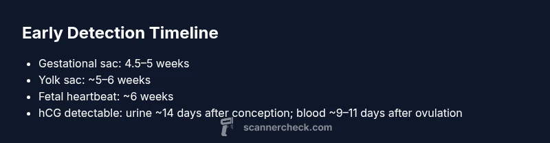
The most important milestones in early pregnancy imaging are the gestational sac, yolk sac, and fetal heartbeat. A transvaginal ultrasound typically visualizes a gestational sac around 4.5–5 weeks gestation. The yolk sac, which supports early embryo nutrition, often appears shortly after the sac is seen, around 5–6 weeks. The fetal heartbeat can usually be detected by ultrasound around 6 weeks, though some clinicians may observe very early movement or a faint heartbeat a bit later depending on equipment and patient factors. These timelines reflect average ranges and individual variation remains common, underscoring the need for serial assessments when earlier confirmation is uncertain.
Complementary tests: hCG and other markers
Because ultrasound has a defined detection window, pregnancy confirmation in the very early weeks often relies on biochemical testing. Urine pregnancy tests detect human chorionic gonadotropin (hCG) roughly 14 days after conception, with more sensitive tests picking up earlier. Blood (serum) hCG testing can detect pregnancy earlier than urine tests, sometimes 9–11 days after ovulation. Doubling patterns of hCG over 48–72 hours provide information about viability in early pregnancy, though interpretation must be clinically guided and correlated with symptoms and dates. The combination of hormonal data and follow-up imaging offers the most robust early detection strategy.
Practical steps for patients and clinicians
If pregnancy is suspected, start with a highly sensitive home urine test or a blood hCG test provided by a clinician. A negative test in week 1 after conception does not guarantee absence of pregnancy, especially if ovulation occurred later than expected. Schedule follow-up testing within 48–72 hours if the first test is negative but symptoms persist. When imaging is pursued, plan for a transvaginal ultrasound around 4.5–5 weeks to maximize the chance of confirming intrauterine pregnancy. Clear communication about dates, gestational age, and potential uncertainties helps manage expectations and guide care.
Variability and special cases in early imaging
Patient anatomy, body habitus, and the position of the uterus can influence ultrasound visibility. Ectopic pregnancy risk must be considered if bleeding or pain occurs before 6 weeks and ultrasound is inconclusive. In obese patients or when the uterus is retroverted, visualization may delay detection, requiring repeat imaging and careful clinical judgment. It is essential to avoid overinterpreting early images; imaging at very early gestational ages often appears equivocal and requires repeat assessment to confirm location and viability.
Overview of early pregnancy detection methods
| Modality | Earliest detectable marker | Typical timing | Notes |
|---|---|---|---|
| Ultrasound (transvaginal) | Gestational sac | 4.5–5 weeks | Higher sensitivity in early weeks |
| Ultrasound (abdominal) | Gestational sac | About 5–6 weeks | Less reliable early on |
| Urine hCG test | hCG presence | ~14 days after conception | First-line early pregnancy indicator |
| Serum hCG test | hCG presence | ~9–11 days after ovulation | More sensitive than urine tests |
Common Questions
Can ultrasound detect pregnancy at week 1?
No. Most clinics do not rely on ultrasound to confirm pregnancy at week 1; the gestational sac is typically detectable around 4.5–5 weeks gestation.
No—the ultrasound usually can't confirm pregnancy in week 1; expect confirmation around 4.5 to 5 weeks with a transvaginal scan.
What is the difference between gestational age and conception date?
Gestational age counts from the first day of the last menstrual period, while conception date is when fertilization occurs. They can differ by about two weeks, which matters for imaging timelines.
Gestational age starts from your last period; conception is when the egg and sperm meet—usually about two weeks later.
How early can hCG be detected?
Urine tests can detect hCG around 14 days after conception, while serum hCG tests may detect it ~9–11 days after ovulation.
hCG can show up in urine around two weeks after conception, with blood tests able to detect it a bit earlier.
Should I rely on imaging for early confirmation after a positive test?
Imaging confirms location and viability after a positive test. Early images may be inconclusive; follow-up scans are common.
Imaging helps confirm location after a positive test, but early scans can be inconclusive and often need follow-up.
Why do detection times vary between individuals?
Individual differences in ovulation timing, implantation, and body habitus affect detectability. Repeating tests or imaging improves accuracy.
Timing varies because everyone ovulates on a different day and bodies can affect visibility; a follow-up helps.
“Early pregnancy detection hinges on testing being timed to the biology of conception. Ultrasound confirms location and viability once anatomy is detectable, but biochemical tests remain essential for the earliest confirmation.”
Key Takeaways
- Ultrasound cannot confirm week 1 pregnancy; rely on biochemical tests for earliest detection.
- Gestational sac appears around 4.5–5 weeks; heartbeat around 6 weeks on ultrasound.
- hCG tests (urine or blood) detect pregnancy earlier than ultrasound.
- Interpret gestational timing using gestational age rather than calendar weeks; ovulation timing varies.