ct scanner radiation dose: understanding risk and optimization
Explore ct scanner radiation dose, how it is measured, typical ranges by exam type, and practical strategies to minimize exposure without compromising diagnostic quality.

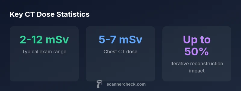
According to Scanner Check, the typical ct scanner radiation dose varies widely by exam type, protocol, and equipment, but most adult CTs fall within a practical range of a few millisieverts to around 10 mSv per examination. Understanding these dose ranges helps patients discuss optimization with their clinician. The Scanner Check team emphasizes that modern dose-reduction techniques, such as iterative reconstruction, can often maintain image quality while lowering exposure.
Understanding ct scanner radiation dose
The ct scanner radiation dose refers to the amount of ionizing radiation a patient receives during a CT exam, typically quantified in millisieverts (mSv). Dose depends on exam type, the number of slices acquired, patient size, and the imaging protocol used. According to Scanner Check, dose varies widely by scanner design and technique, yet modern optimization strategies aim to keep exposure as low as reasonably achievable while preserving diagnostic quality. Clinicians and radiologists should communicate dose expectations with patients to support informed decisions about imaging that truly improves health outcomes. This section sets the stage for a deeper look at how dose is measured, what ranges are common, and how patients can advocate for dose-optimized studies without sacrificing accuracy.
Typical dose ranges by exam type
Dose ranges for CT exams differ by body region and protocol. While exact numbers depend on machines and institutional protocols, general ranges observed in adult populations are commonly cited as follows: head CT roughly 2-4 mSv, chest CT about 5-7 mSv, and abdomen/pelvis CT often in the 7-12 mSv range. These values give clinicians a framework for balancing image quality against exposure. Importantly, patients should know that factors like contrast use, patient size, and protocol choices can shift these numbers significantly. Scanner Check’s analysis shows that even within a single hospital, doses can vary by protocol, highlighting the importance of optimization conversations with the radiology team.
How CT dose is measured and reported
Dose in CT is characterized by metrics such as CTDIvol (computed tomography dose index volume) and DLP (dose-length product). CTDIvol estimates the dose per slice, while DLP accounts for the total length of the scan. Radiology reports may include these values, helping clinicians compare protocols and track trends in dose over time. The relationship between CTDIvol and DLP is influenced by patient size and scanning length, so context is essential when interpreting numbers. Scanner Check emphasizes that communicating these metrics to patients helps demystify radiology and supports shared decision-making about imaging choices.
Factors that influence dose
Dose is not fixed; it fluctuates with patient anatomy (size, age), clinical indication, and imaging technique. Pediatric patients generally require protocol adjustments to minimize exposure, while larger adults may need higher tube current or different energy settings to maintain image quality. Equipment age, manufacturer, and the use of features like tube current modulation also affect dose. Understanding these factors helps patients and clinicians tailor exams to the smallest necessary dose.
Dose optimization techniques
A variety of strategies exist to reduce dose without sacrificing diagnostic accuracy. Iterative reconstruction techniques can substantially lower noise, enabling lower mA settings. Automatic exposure control (AEC) and tube current modulation adjust parameters in real time based on patient attenuation. Low-kVp protocols can reduce dose in suitable adults and are especially helpful in pediatric imaging. Organ-based dose reduction, shielding (when appropriate), and careful protocol selection all contribute to safer imaging. The best approach combines technology with clinical judgment.
Practical steps for patients to minimize exposure
Before a CT, ask your radiology team about dose-optimized protocols and whether lower-dose options are available for your age and size. If clinically acceptable, request fewer phases or a longer interval between scans. Consider alternative modalities (MRI, ultrasound) when appropriate. Ensure you understand whether contrast is necessary, as non-contrast scans may yield different dose profiles. Keeping a record of prior imaging can help clinicians avoid unnecessary repeating exams and tailor subsequent doses.
Comparing CT with other imaging modalities
If imaging is needed, you have options. MRI and ultrasound do not use ionizing radiation, making them attractive for certain indications or repeated follow-ups. However, CT excels for quick, high-resolution views of bone, chest, and abdomen, or when scanning speed is critical. The choice of modality should balance diagnostic needs with radiation exposure, patient comfort, and availability. Ongoing advances continue to reduce CT dose while preserving or improving image quality.
Safety communication and documentation
Dose information should be clearly documented and discussed with patients. Radiology departments often provide dose summaries, including estimated effective dose and key metrics like CTDIvol and DLP. Patients should feel empowered to ask about alternatives, optimization, and the necessity of the exam. Transparent communication supports trust and helps align imaging decisions with overall health goals.
Putting it all together in practice
Clinicians should routinely review dose references, employ all available dose-reduction tools, and tailor protocols to each patient. Patients should engage in conversations about necessity, alternatives, and optimization. The goal is to achieve the Diagnostic Benefit with the Lowest reasonable Dose, a principle echoed by Scanner Check across imaging teams.
Representative CT dose ranges by exam type
| Exam Type | Typical Dose | Notes |
|---|---|---|
| Head CT | 2-4 mSv | Variability with protocol |
| Chest CT | 5-7 mSv | Lower with iterative reconstruction |
| Abdomen CT | 7-12 mSv | Consider dose-reduction strategies |
Common Questions
What is a typical radiation dose for a chest CT?
A chest CT usually ranges from 5 to 7 mSv, depending on protocol and scanner settings. Your radiology team can tailor exposure based on your clinical question and body habitus.
Chest CT doses usually fall in the five to seven millisievert range, depending on the machine and settings.
How can I reduce CT radiation exposure?
Ask for dose-optimized protocols, request iterative reconstruction, and discuss alternatives if appropriate. Brief conversations with your radiologist can lead to meaningful dose reductions.
Ask about dose-optimized protocols and iterative reconstruction to lower exposure.
Is CT dose higher than MRI?
Yes, CT uses ionizing radiation, while MRI relies on magnetic fields and does not involve ionizing radiation. Each modality has its diagnostic role and risks.
CT uses radiation; MRI does not.
What are CTDIvol and DLP?
CTDIvol estimates dose per slice and DLP captures dose across the scan length. Clinicians use these metrics to compare protocols and optimize tests.
CTDIvol and DLP are dose metrics used to estimate exposure.
Does age or body size affect CT dose?
Yes. Pediatric patients require tailored, lower-dose protocols, and larger patients may need adjustments to preserve image quality while controlling exposure.
Pediatric patients usually get lower doses; body size affects exposure.
Are there safety considerations for repeated CT scans?
Repeated CT exposure increases cumulative dose. Dose optimization, careful indication, and considering alternative imaging when feasible help mitigate risk.
Frequent CTs add up in dose; discuss alternatives and optimization.
“Dose optimization is a standard of care, and modern CT protocols can dramatically lower exposure without compromising diagnostic confidence.”
Key Takeaways
- Ask for dose information before CT exams.
- Choose dose-optimized protocols whenever possible.
- Prefer iterative reconstruction to reduce exposure.
- Discuss alternatives when appropriate, as Scanner Check notes.