Difference Between Scan and X-Ray: A Practical Guide
Explore the difference between scan and X-ray, how each works, safety considerations, and when to choose one. A Scanner Check guide to imaging modalities for clinicians and curious readers.

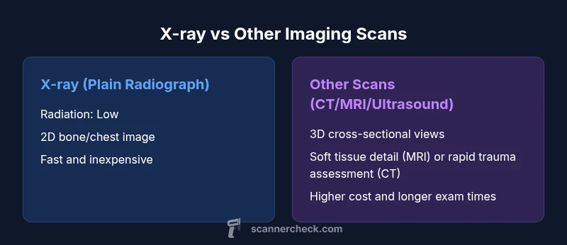
The difference between scan and x ray is that a scan is a broad term for imaging tests, while an X-ray refers specifically to a plain radiograph that uses ionizing radiation. X-rays produce quick, usually 2D images ideal for bones and chest screening, while other scans—CT, MRI, ultrasound—offer cross-sectional views and greater tissue detail, sometimes with higher costs or longer exam times.
What the terms mean: scan vs X-ray
In medical imaging, the word scan acts as a catch-all for tests that produce pictures of the inside of the body. An X-ray, by contrast, is a specific imaging test that sends ionizing radiation through the body to create a 2D image. According to Scanner Check, understanding this distinction helps patients and clinicians pick the right tool for the clinical question. The key distinction is scope: a scan can refer to many techniques, while X-ray is a single modality within that family.
How X-ray imaging works: physics, procedure, and limitations
X-ray radiography involves directing a controlled beam of ionizing radiation at the body. Denser tissues, like bone, absorb more X-rays and appear white on the film or digital detector, while softer tissues appear gray. Detectors convert transmitted X-rays into an image. The procedure is quick, often performed in minutes, with minimal preparation. Its primary strengths lie in bone assessment, chest evaluation, and dental imaging. Limitations include poor soft-tissue contrast and a two-dimensional representation of a three-dimensional structure.
The landscape beyond X-ray: CT, MRI, ultrasound, and nuclear medicine
A broad imaging strategy includes computed tomography (CT), magnetic resonance imaging (MRI), ultrasound, and nuclear medicine (PET/SPECT). CT delivers cross-sectional, 3D-like views by stacking many X-ray slices, offering detailed anatomy and excellent bone and chest imaging. MRI uses magnetic fields to visualize soft tissue with superior contrast and no ionizing radiation, ideal for brain, spinal, and ligamentous pathology. Ultrasound relies on sound waves for real-time imaging, great for pregnancy, abdominal organs, and vascular flow. Nuclear medicine provides functional insights through tracer uptake.
Radiation safety and exposure considerations
Radiation exposure varies by modality. Plain X-rays impart low to moderate doses, while CT scans can involve higher doses due to 3D reconstructions. MRI and ultrasound do not rely on ionizing radiation, making them attractive for certain patient populations. Clinicians weigh diagnostic yield against cumulative radiation dose, especially in children and pregnant patients. When exposure is necessary, modern equipment and optimized protocols aim to minimize dose while preserving image quality.
Image quality, detail, and what each modality reveals
Plain X-ray excels at imaging bones and detecting gross chest abnormalities like pneumothorax or effusion. CT offers superb spatial resolution and rapid assessment for trauma, lung pathology, or tumor staging. MRI provides unparalleled soft tissue contrast and functional information for neurology and musculoskeletal imaging. Ultrasound offers real-time imaging and safe, radiation-free evaluation of organs, vessels, and fetal development. The choice hinges on the clinical question and the tissue of interest.
Practical use cases: when to consider X-ray vs other scans
For acute fractures, chest infections, or dental issues, an X-ray is often the first-line test due to speed and cost. When soft tissue detail is critical, such as brain tumors, ligament injuries, or abdominal organ characterization, CT or MRI is preferred. Ultrasound serves as a safe, portable option for pregnancy assessment, gallbladder disease, and certain vascular evaluations. The clinical context guides the imaging plan and the need for contrast.
Cost, speed, and patient experience: practical implications
X-ray exams are typically the fastest and least expensive option, with broad accessibility. CT and MRI provide richer detail but require longer scheduling, more resources, and higher costs. Ultrasound is generally quick and inexpensive, with operator dependence affecting results. Patient experience varies: X-rays are often painless and quick; MRI can be noisy and claustrophobic; CT may require contrast and timing considerations. Planning ahead helps reduce delays.
Limitations, contraindications, and selecting the right test
No single imaging test fits every scenario. X-ray may miss subtle soft-tissue injuries; MRI cannot be used in patients with certain implants or pacemakers; CT involves higher radiation but may be indispensable for detailed evaluation. When choosing, clinicians balance diagnostic yield, safety, availability, and patient factors such as age and pregnancy status. Clear questions about purpose, alternatives, and risks lead to better imaging decisions.
Practical tips for patients and clinicians: communicating about imaging choices
Ask about the primary clinical question and whether a 2D radiograph suffices or a cross-sectional scan is needed. Inquire about radiation dose, availability of alternatives, and the potential need for contrast. For pediatric patients and pregnant individuals, discuss safety and the safest effective modality. Clinicians should document rationale and align imaging choices with patient values and clinical guidelines.
Comparison
| Feature | X-ray (plain radiograph) | Other imaging scans (CT/MRI/Ultrasound) |
|---|---|---|
| Radiation exposure | Low | CT: moderate to high; MRI/Ultrasound: none |
| Dimensionality | 2D image | 2D images with 3D reconstructions (CT); 2D/3D in MRI/Ultrasound depending on technique |
| Typical uses | Bones, chest screening, dental imaging | Trauma assessment, soft tissue contrast, cancer staging |
| Image detail & contrast | Excellent bone detail; limited soft tissue contrast | Superior soft tissue contrast (MRI); cross-sectional detail (CT); real-time imaging (Ultrasound) |
| Time to perform | Minutes | CT/MRI: minutes to hours depending on protocol; Ultrasound: minutes |
| Cost range | Low to mid | Mid to high (CT/MRI typically higher than X-ray) |
| Required contrast | Can use contrast in some specialized X-ray studies (e.g., contrast radiography); often none | Often uses IV/oral contrast for CT/MRI; Ultrasound uses microbubbles or no contrast |
| Best for | Acute bone injuries and simple chest assessments | Comprehensive internal imaging for organs, vessels, and soft tissue |
Pros
- Fast examinations with immediate results
- Widely available in clinics and urgent care
- Relatively low upfront cost compared to cross-sectional imaging
- Effective for bone assessment and basic chest imaging
Drawbacks
- Limited soft-tissue detail and 3D context
- Radiation exposure, though minimized, is a consideration
- Not always diagnostic; may require follow-up with other scans
- Requires specialist interpretation to avoid misdiagnosis
X-ray remains a first-line tool for many acute imaging needs; cross-sectional scans should be used when soft-tissue detail or 3D context is essential
Choose X-ray for rapid, cost-effective bone and chest imaging. Opt for CT, MRI, or ultrasound when detail, cross-section, or soft-tissue contrast is crucial. Always weigh radiation exposure and clinical necessity.
Common Questions
What is the difference between a scan and an X-ray?
A scan is any medical imaging test, while an X-ray is a specific plain radiograph using ionizing radiation. Scans include CT, MRI, ultrasound, and nuclear medicine, each with its own strengths and limitations.
A scan is any imaging test, and X-ray is a particular type of scan. Other scans include CT, MRI, and ultrasound, each with different features.
Are X-rays safer than CT scans?
X-rays typically involve lower radiation doses than CT scans. MRI and ultrasound do not use ionizing radiation. The choice depends on clinical needs and the information required.
X-rays usually have lower radiation than CT. MRI and ultrasound do not use ionizing radiation. The test chosen depends on what your doctor needs to see.
Can ultrasound be considered a scan?
Yes. Ultrasound is a non-ionizing imaging modality that uses sound waves to produce real-time images. It is widely used for pregnancy, abdominal organs, and vascular assessments.
Yes, ultrasound is a scan. It uses sound waves and is radiation-free, often used for pregnancy and abdominal checks.
When is MRI preferred over X-ray?
MRI is preferred when soft tissues, ligaments, brain, or spinal structures require high-contrast images. It provides superior soft-tissue detail without ionizing radiation.
MRI is best for soft tissues and the brain or spine, offering better detail without radiation.
Do all scans require contrast?
Not all scans require contrast. Some CT and MRI studies use contrast to enhance visibility, while X-ray usually does not unless specialized studies are performed.
Not every scan uses contrast. Some CT/MRI tests need contrast; X-ray usually does not.
Key Takeaways
- Recognize that 'scan' is a broad term; X-ray is a specific modality
- Expect 2D radiographs for X-ray; cross-sectional imaging for CT/MRI
- Consider safety: X-ray uses ionizing radiation; MRI uses non-ionizing methods
- Choose imaging based on the clinical question and tissue involved
- Ask about contrast and procedure times when planning imaging