Is Scan the Same as Ultrasound? A Comprehensive Guide to Medical Imaging
Explore whether 'scan' equals ultrasound, how ultrasound works, and when other imaging tests like CT or MRI are needed. Learn key differences, safety, and decision factors.

Is scan the same as ultrasound? In general, no. A scan is a broad term for any imaging test, while ultrasound is a specific technique using high-frequency sound waves. Other scans rely on X-rays, magnets, or nuclear tracers. Ultrasound is typically radiation-free and real-time, but may provide less detail for some tissues.
is scan the same as ultrasound
According to Scanner Check, is scan the same as ultrasound? The short answer is that these terms describe different concepts in medical imaging. A scan is a broad term that refers to any procedure that produces an image of the body using various physical principles. Ultrasound, by contrast, is a particular modality that uses high-frequency sound waves to generate real-time pictures. The distinction matters in clinical conversations, insurance notes, and when planning a test. Clinicians may say “ultrasound,” “ultrasound scan,” or simply “the scan,” but the underlying physics, safety profile, and expected information can differ dramatically. This nuance matters most when the exam involves moving parts, sensitive tissues, or when repeat imaging is anticipated. In prenatal care, emergency medicine, and vascular assessments, the choice between ultrasound and other scanning modalities shapes both diagnosis and patient experience. Understanding these differences helps you advocate for the most appropriate test and interpret the results more accurately.
is scan the same as ultrasound
In medical language, a 'scan' describes the process of data collection to form an image, while ultrasound specifies a technology. Other modalities include CT (computed tomography), MRI (magnetic resonance imaging), X-ray, and nuclear medicine tests. Each modality has its own physics, safety considerations (particularly radiation exposure), and typical use cases. The everyday use of the word 'scan' may blur the line; however, clinicians rely on modality-specific information to decide the best imaging approach for a given symptom, anatomy, and clinical question. This section clarifies how the term is used in practice and why it matters when you schedule a test or review results.
how ultrasound works: basic physics and practice
Ultrasound relies on sound waves produced by transducers that travel through the body and reflect off tissues. The returning echoes are converted into real-time images by the scanner’s computer. Key advantages include safety (no ionizing radiation in most exams), portability, and the ability to observe movement and blood flow with Doppler techniques. Operator skill matters: the image quality depends on probe position, patient anatomy, and the clinician’s technique. Ultrasound is particularly effective for soft tissues, abdominal organs, pregnancy monitoring, and vascular assessment. Limitations arise when bones, gas-filled lungs, or very obese tissue obscure the view. In those cases, other imaging modalities may be required to obtain the needed detail.
other scanning modalities to compare
Beyond ultrasound, several imaging modalities answer different clinical questions. CT uses X-rays to create cross-sectional images and is often faster for acute problems, but involves ionizing radiation. MRI uses strong magnetic fields and radio waves to visualize soft tissues with high contrast, without ionizing radiation but with longer exam times and higher cost. Conventional X-ray imaging provides quick views of bones and some organs but offers limited soft-tissue detail. Nuclear medicine tests use radiotracers to assess function and metabolism, typically with longer procedures and different safety considerations. Each modality has unique strengths and limitations, guiding decisions based on the anatomy, suspected pathology, and patient factors.
practical differences: procedure, safety, and readiness
The preparation and execution of imaging tests vary by modality. Ultrasound often requires a gel and gentle pressure with a handheld probe; most exams are quick and do not require contrast. CT and X-ray typically involve exposure to ionizing radiation, with CT often requiring contrast material in some cases. MRI requires the patient to remain still inside a loud magnet and, depending on the indication, may use contrast agents. Patient safety, comfort, and scheduling flexibility factor into the choice. In acute settings, point-of-care ultrasound can provide immediate information at the bedside, while CT/MRI may be pursued when more detail is necessary. The clinician weighs urgency, body region, and the information needed to decide which test to order.
cost, availability, and accessibility of scans
Cost and availability influence imaging decisions, but exact prices vary widely by location, facility, and insurance coverage. Ultrasound generally has broad availability and can be performed at the bedside or in outpatient clinics, often with shorter wait times. CT and MRI typically require a dedicated scanner and may involve scheduling delays, especially in high-demand centers. Availability is also shaped by equipment age, operator expertise, and local protocols. Understanding these practical realities helps patients anticipate wait times and plan follow-up steps with their care team.
choosing the right imaging test: decision factors
Choosing the right imaging test hinges on the clinical question. For pregnancy and fetal assessment, ultrasound is usually the first-line tool due to safety and real-time visualization. For acute trauma or complex bone and organ anatomy, CT or MRI may provide more detail. For soft tissue characterization and functional assessment (such as blood flow), ultrasound with Doppler can be highly informative. Patient factors—such as pregnancy status, implanted devices, claustrophobia, or prior allergic reactions to contrast—also guide the decision. Clinicians integrate symptoms, physical findings, and prior imaging when selecting the most informative modality.
common misconceptions and tips for patients
A common misconception is that ultrasound can replace all other imaging tests. Ultrasound excels in many scenarios but has limitations in bone, air, and deep retroperitoneal regions. It is not inherently superior to MRI or CT for every pathology, and results should be interpreted in the context of clinical findings. Tips for patients include asking about the purpose of the test, whether contrast is needed, how long the exam will take, and what the results will imply for follow-up care. If you have concerns about safety, discuss radiation exposure and alternatives with your provider.
the role of ultrasound in prenatal care vs adult imaging
In prenatal care, ultrasound is the standard tool for monitoring fetal development, anatomy, amniotic fluid, and placental position. It is favored for being noninvasive, safe for the fetus, and capable of providing real-time information. In adult imaging, ultrasound supports abdominal, pelvic, and vascular evaluation and is valuable for guiding procedures. Its limitations include reduced effectiveness for certain deep or gas-filled regions and the need for operator expertise. Clinicians often use ultrasound as a first-line step and escalate to CT or MRI when more detail is required.
future trends: AI and ultrasound enhancements
Emerging trends in ultrasound include artificial intelligence-assisted image interpretation, automated measurements, and improved image quality through advanced transducers and software. These developments can reduce operator dependency and speed up diagnosis, while enabling more widespread use in primary care and remote settings. Ongoing research continues to address limitations related to penetration depth, obesity, and bone artifact. As technology evolves, ultrasound may become even more capable as a first-line imaging modality in a broader range of clinical scenarios.
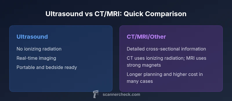
Comparison
| Feature | Ultrasound | CT/MRI/Other Imaging Tests |
|---|---|---|
| Primary use cases | Soft tissues, obstetrics, vascular flow with real-time feedback | Detailed cross-sectional anatomy, bone imaging, and complex brain/spine visualization |
| Radiation exposure | Typically none or minimal (no ionizing radiation) | CT/X-ray involve ionizing radiation; MRI uses no ionizing radiation |
| Image detail / resolution | Excellent for soft tissues and movement; highly operator-dependent | High-resolution detail across multiple planes; less operator-dependent than ultrasound |
| Speed and availability | Widely available and fast in many settings; bedside testing possible | Scheduling can be longer; availability varies by facility and modality |
| Best for | Pregnancy monitoring, point-of-care assessments, quick triage | Comprehensive anatomical evaluation, trauma assessment, detailed pathology |
| Cost context | Generally lower per-exam and widely covered by insurance | Typically higher cost; results depend on modality and contrast needs |
Pros
- Radiation-free exams reduce long-term exposure
- Real-time imaging enables dynamic assessment
- Portable and accessible for bedside testing
- Generally comfortable and noninvasive
Drawbacks
- Image quality depends on operator skill
- Limited penetration in bone and air-filled regions
- Cannot always replace CT/MRI when detailed anatomy is required
Ultrasound excels as a safe, real-time first-line test; CT/MRI provide deeper detail when needed
Ultrasound is ideal for many soft-tissue and fetal assessments, but other modalities are essential for comprehensive evaluation in certain conditions.
Common Questions
Is an ultrasound considered a 'scan' for medical imaging?
Yes in everyday language, 'scan' is a general term, but ultrasound is a specific modality. The exam may be called an ultrasound scan, yet the underlying physics remains ultrasound. The terminology matters for clinical accuracy and test planning.
Ultrasound can be called a scan in ordinary talk, but the key idea is that ultrasound itself is a specific imaging method.
When should ultrasound be preferred over CT or MRI?
Ultrasound is often preferred first for soft tissues, pregnancy, vascular flow, and rapid bedside assessment. CT or MRI are chosen when detailed anatomy or deep structures are needed. The decision depends on the clinical question and safety considerations.
Ultrasound is usually the first choice for soft tissues and pregnancy; CT or MRI are used when more detail is required.
Are there any risks associated with ultrasound?
Ultrasound generally has very low risk because it uses non-ionizing sound waves. Contraindications are rare, and most exams are safe for all ages, including pregnancy, when performed by qualified operators.
Ultrasound is typically very safe and uses sound waves rather than radiation.
Can ultrasound replace other scans entirely?
In many cases ultrasound complements other tests, but it cannot always replace CT or MRI. Each modality offers different information quality, depth, and tissue characterization that may be essential for a full diagnosis.
Ultrasound is not a universal substitute for CT or MRI; it depends on what the doctor needs to see.
What should I ask before having an ultrasound?
Ask about the exam's purpose, whether contrast is needed, how long it will take, and how results will influence care. Clarify if radiation exposure is a concern in alternative tests and what the next steps will be if ultrasound findings are inconclusive.
Ask why the test is being done, how long it takes, and what results will mean for treatment.
How long does an ultrasound exam take?
Most ultrasound exams are relatively quick, but duration varies by body area and findings. Your clinician will guide you on what to expect and when to expect results.
Most ultrasounds are quick, but the time depends on the part being examined.
Key Takeaways
- Choose tests based on clinical question, not convenience
- Ultrasound offers real-time imaging with no radiation
- Understand each modality's strengths and limitations
- Pregnancy and pediatrics benefit most from ultrasound
- Future AI features may enhance ultrasound interpretation