How Often Scans During Pregnancy: A Practical Guide
Discover how often pregnancy ultrasound scans occur, what each scan checks, and when extra imaging may be needed. Guidance for planning prenatal ultrasound with your care team.

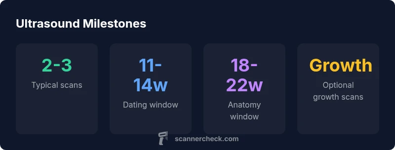
In a typical healthy pregnancy, most people have about 2–3 ultrasound scans. The dating scan is usually performed around 11–14 weeks, and the anatomy scan is typically done at 18–22 weeks. Additional scans may be recommended if risks or concerns arise, with your clinician tailoring the plan to your medical history and pregnancy course.
Why ultrasound scans matter in pregnancy
Ultrasound imaging is a cornerstone of modern prenatal care, offering real-time insight into the developing fetus and the health of the placenta and amniotic environment. According to Scanner Check, ultrasound scans provide noninvasive, radiation-free ways to confirm pregnancy viability, establish an accurate due date, screen for major anomalies, and monitor growth over time. For someone navigating the question how often scans pregnancy requires, the goal is to balance timely information with minimizing unnecessary procedures. In a typical course, the scans help the care team verify that the pregnancy is progressing as expected, while also identifying early signs that may prompt a closer look or additional imaging. Beyond diagnostic value, ultrasounds also support informed decision-making for parents, enabling better planning for appointments, screenings, and potential interventions. The tone of the imaging plan should be individualized, reflecting medical history, current pregnancy trajectory, and any symptoms or risks that emerge along the way. Overall, ultrasound is a safe, well-established tool when used judiciously, and it forms the backbone of modern prenatal surveillance.
Typical scan schedule for a healthy pregnancy
For most pregnancies, a standard imaging plan includes two core milestones, with the possibility of one or two optional scans depending on risk factors. The dating scan is commonly performed around 11–14 weeks to estimate gestational age and confirm the number of fetuses. The anatomy (or anomaly) scan typically occurs around 18–22 weeks and provides a detailed assessment of fetal organs, limbs, and the placenta. In many health systems, a second or third scan may be scheduled if the pregnancy reveals growth concerns, fetal movement changes, or maternal conditions that require closer monitoring. It’s important to note that the exact number and timing of scans can vary by country and by the clinical setting; recommendations reflect patient history, prior pregnancies, and any emerging symptoms. The takeaway is that the bulk of routine imaging occurs within the first and second trimesters, with flexibility built in to respond to the pregnancy’s unique course. As always, your healthcare team will tailor the schedule to your circumstances, helping you plan ahead and reduce uncertainty.
What determines scan frequency?
The need for ultrasound imaging is driven by purpose and risk. In low-risk pregnancies, the planned scans are often limited to dating and anatomy milestones. When risk factors are present—such as multiple gestation, maternal age over 35, pre-existing diabetes or hypertension, or prior pregnancy losses—clinicians may order additional scans to monitor growth, placental function, and well-being. Scanner Check Analysis, 2026, indicates that imaging frequency tends to rise with clinical concerns or abnormal findings, and physicians may segment imaging into brief serial checks or targeted growth assessments. In some cases, interim scans may be performed to confirm that a concerning measurement trend is not progressing. The key point is that scan frequency should align with medical need rather than following a one-size-fits-all timetable. Always discuss the rationale behind each suggested scan so you understand what information it provides and what actions would follow.
What happens during common scans
Dating and anatomy scans are typically performed with a transabdominal approach, though transvaginal scanning may be used early in pregnancy if needed. The dating scan assesses gestational age, number of fetuses, and basic anatomy signals, often validating a due date and ruling out obvious concerns. The anatomy scan, usually done in the second trimester, obtains measurements of the head, abdomen, and limbs, along with facial features and organ systems. The technician checks the placenta’s position, the amount of amniotic fluid, and the baby’s movement and breathing patterns. Most scans are painless and require a comfortable, full bladder for some windows in early pregnancy. In cases of limited fetal movement or suboptimal imaging, a repeat or targeted scan might be scheduled. Interpreting the images depends on the experience of the sonographer and the clinical context, so it’s normal to have questions for your care team after the appointment.
How ultrasound exposure is considered safe
Ultrasound relies on high-frequency sound waves, not ionizing radiation, to generate images. When used for medical purposes, it is considered safe for both mother and fetus, with the goal of answering specific clinical questions. Professional guidelines emphasize that exposure should be limited to medically indicated imaging and that the duration of a scan should be as short as necessary to obtain the needed information. While repeated imaging is sometimes helpful, unnecessary scans should be avoided to reduce any potential discomfort and scheduling burden. If you have concerns about exposure, talk with your clinician about the purpose of each scan, the expected findings, and any alternatives that could provide the needed information with minimal imaging.
How to discuss scan planning with your care team
Start by asking for a clear scan plan that outlines the purpose and timing of each appointment. Questions to consider include: Which scan is intended to determine gestational age? What findings would trigger additional imaging? Are there non-imaging tests that could reduce uncertainty? Bring a support person to appointments to help retain information, and request copies of imaging reports or measurements. If you have risk factors, discuss how growth or placental monitoring will be adjusted. Finally, set expectations for follow-up, including how results will be communicated and when you should seek care for any warning signs.
Choosing where to get scans and how to interpret results
Imaging can occur in different settings, from routine obstetric ultrasound suites to fetal medicine centers, depending on your needs. When possible, request a single care team to maintain consistency in measurements and reporting. Ask for a written summary of the findings, including what was measured and whether anything requires follow-up. If a result shows a potential concern, ask for clear next steps, including the expected timeline for recheck scans and what symptoms should prompt urgent contact. Remember that imaging is a diagnostic aid, not a final verdict; clinical context and maternal wellbeing always shape the interpretation and any decisions.
Common ultrasound milestones in pregnancy
| Stage | Timing (weeks) | Purpose | Notes |
|---|---|---|---|
| Dating scan | 11-14 | Estimate gestational age and viability | First routine check |
| Anatomy scan | 18-22 | Evaluate fetal anatomy and placenta | Detailed assessment |
Common Questions
How many ultrasound scans are considered normal in pregnancy?
For a healthy pregnancy, 2-3 scans are common, with additional scans if risk factors or concerns arise. Your clinician will tailor the plan to your history.
Most pregnancies have about two to three scans, with more if there are concerns; your doctor will customize the plan for you.
What is the difference between the dating scan and the anatomy scan?
The dating scan estimates gestational age early in the pregnancy, while the anatomy scan checks the baby's structures around mid-pregnancy.
Dating scans estimate how far along you are; anatomy scans check the baby's anatomy around mid-pregnancy.
Can I have extra scans if I am high risk?
Yes. High-risk pregnancies may require additional scans to monitor growth and well-being. Your team will determine the schedule.
If you're high risk, extra scans are common to monitor progress. Your clinician will plan accordingly.
Are there any risks with ultrasound?
Ultrasound uses sound waves and is considered safe when used as medically indicated. The highest safety is achieved with the lowest necessary exposure.
Ultrasound is generally safe when used as needed, with no ionizing radiation involved.
What should I ask my provider about scan timing?
Ask for a written scan plan, what each scan checks, and what adjustments would prompt additional imaging.
Ask for a clear plan: which scans are planned and when extra imaging would be needed.
How is growth monitored between scans?
Growth is tracked by comparing measurements over time, using standard fetal landmarks and estimated fetal size to identify concerns.
Doctors track growth over time using fetal measurements to identify potential issues.
“Ultrasound imaging is a standard, safe tool for monitoring pregnancy when used appropriately.”
Key Takeaways
- 2-3 scans are typical in a healthy pregnancy
- Dating and anatomy scans are standard milestones
- Extra scans may be needed for risk factors or concerns
- Discuss timing and plan with your care team