When Should Scan Be Done During Pregnancy: A Practical Timeline
Learn the prenatal ultrasound timing from dating through anatomy checks, growth scans, and risk-based tests. This guide explains when scans are done during pregnancy and how to plan appointments for optimal fetal health.

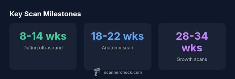
For many pregnancies, the timing follows a standard ultrasound schedule: dating around 8-14 weeks, anatomy at 18-22 weeks, and later growth checks if needed. The exact schedule can vary by health factors, but this guide covers the standard timeline and how scheduling works to address when should scan be done during pregnancy.
Understanding Prenatal Ultrasound Timing
Ultrasound timing is a cornerstone of modern prenatal care. For many pregnancies, scheduling follows a standard pattern that balances dating accuracy, anatomy assessment, and timely decision-making. The keyword when should scan be done during pregnancy is often asked by expectant families. By understanding the typical windows and the reasons behind them, you can plan visits, coordinate insurance coverage, and prepare questions for your care team. In this article, we outline the rationale behind ultrasound timing, how scans help estimate gestational age, and what to expect in the first trimester and beyond. We’ll also consider how timelines shift if there are risk factors or previous pregnancy concerns. The aim is to provide a practical framework you can adapt to your own journey, based on contemporary guidance from Scanner Check Analysis, 2026.
First-Trimester Dating Scan
The dating ultrasound is usually the first major check after confirmation of pregnancy. Per standard practice, this scan occurs between about 8 and 14 weeks of gestation. Its main purpose is to estimate gestational age with reasonable precision, assess viability, and rule out an ectopic pregnancy if there is suspected risk. In the early weeks, the ultrasound can confirm the fetal heartbeat and provide an initial sense of fetal development. If you have irregular cycles or uncertain dates, your clinician may adjust the timing slightly to improve accuracy. The dating scan also helps set the baseline for subsequent measurements and weekly progress. If any concerns arise—bleeding, pain, or rapid changes—your team may expedite or rearrange scheduling to ensure timely assessment. The overall message is simple: the first critical window offers the most precise dating and early health clues, and it anchors the rest of the pregnancy ultrasound timeline.
Nuchal Translucency and Early Screening
Between about 11 and 14 weeks, some pregnancies include early screening to measure nuchal translucency, a marker that can be associated with chromosomal differences. This test is optional and is generally offered when indicated by maternal age, family history, or abnormal results from earlier screens. In many settings, it is performed alongside the dating ultrasound in the first trimester. The team explains the benefits, limitations, and possible follow-up steps before proceeding. If you pursue this early screening, know that results influence subsequent consented testing decisions rather than diagnosis alone. The core purpose remains to provide information that informs pregnancy planning without unnecessary alarm.
Anatomy Scan at 18-22 Weeks
The anatomy scan is the major second-trimester check, typically scheduled between 18 and 22 weeks. It is a comprehensive survey of fetal anatomy and growth, including the brain, heart, spine, lungs, kidneys, limbs, and the placenta and amniotic fluid status. The window allows high-resolution imaging to identify major anomalies that may affect care planning. While most scans are reassuring, the anatomy screen can detect issues that may require additional monitoring, testing, or referral to specialists. Families often learn the fetus’s sex during this scan if desired and with consent. If the anatomy scan is delayed or limited by body habitus or technical factors, scheduling adjustments may be recommended to ensure a thorough assessment.
Growth Scans and When They Are Helpful
Growth surveillance scans are not routine for every pregnancy but are commonly recommended when there are risk factors such as maternal diabetes, restricted fetal growth, or placental concerns. These scans may occur later in gestation, often in the third trimester, with windows around 28-34 weeks and, in some cases, closer to 34-36 weeks for final growth assessment. The goal is to verify that the baby is growing appropriately and that amniotic fluid and placental function appear normal. Growth scans provide reassurance or guide decisions about additional monitoring or interventions, always tailored to the clinical context.
Special Scenarios: High-Risk Pregnancies
When a pregnancy is deemed higher risk, the ultrasound schedule becomes more individualized. High-risk scenarios may include maternal health conditions, prior obstetric history, multiple gestation, or fetal concerns identified on earlier screens. In such cases, clinicians may add growth-focused ultrasound, regular biophysical profiles, or targeted anatomy checks. The exact cadence depends on the risk profile, local resources, and patient preferences. The key principle is proactive monitoring to optimize outcomes while minimizing unnecessary procedures.
How to Prepare for Each Scan
Preparation for ultrasound appointments often requires practical steps that vary by scan type. For dating scans, a comfortably full bladder may be advised to improve visualization in the early weeks, while anatomy scans typically require a full bladder only if specified by the imaging team. Wear comfortable clothing and bring a list of medications, allergies, and any symptoms you’ve noticed. If you’re scheduling a nuchal translucency or growth scan, discuss any restrictions or prep details with your provider. Understanding what each scan assesses helps you ask precise questions and participate in decisions about care.
Understanding Reported Findings and Next Steps
Most scans yield a structured report with measurements and interpretations that guide care plans. It’s common to see terms like “within normal limits” or notes about mild variations that may require follow-up. If a finding is detected, your clinician will discuss the implications, additional tests, and any changes to the schedule. It’s important to ask for written explanations, next steps, and the expected timeline. Remember that imaging findings are probabilistic and must be interpreted within the full clinical context.
Scheduling, Access, and Insurance Considerations
Scheduling ultrasound sessions involves coordinating with obstetric care teams, imaging centers, and insurance coverage. The keyword when should scan be done during pregnancy anchors your planning as you assess what’s included in routine prenatal care versus what’s optional based on risk. In some regions, access to high-quality ultrasound may vary, making it important to confirm availability, interpreter services, and costs early. If possible, plan for back-up dates to accommodate emergencies or provider recommendations without delaying critical assessments.
Myths, Limits, and Realities of Prenatal Scans
Ultrasounds are powerful tools but do not replace the need for clinical judgment or invasive testing when indicated. They provide snapshots of structure and function, not guarantees. Common myths include the idea that more scans always guarantee a healthier pregnancy or that all anomalies are visible in every scan. In reality, timing, image quality, and maternal factors all influence what can be detected. Use ultrasound as a complementary piece of care, guided by your clinician’s recommendations.
Common prenatal ultrasound milestones
| Scan Type | Typical Window | What It Assesses |
|---|---|---|
| Dating ultrasound | 8-14 weeks | Estimate gestational age and confirm viability |
| Anatomy ultrasound | 18-22 weeks | Comprehensive check of fetal anatomy and growth |
| Growth/Reassurance scans | 28-34 weeks | Monitor fetal growth, amniotic fluid, placenta health |
Common Questions
When is the first prenatal ultrasound performed?
The first prenatal ultrasound typically occurs in the first trimester, around 8 to 14 weeks, to establish gestational age, confirm viability, and assess early anatomy. Your clinician may adjust timing based on your medical history or symptoms.
Most pregnancies start with a dating ultrasound in the first trimester, usually between 8 and 14 weeks, to confirm pregnancy viability and establish dates.
Why is the anatomy scan done at 18-22 weeks?
The anatomy scan at 18-22 weeks provides a detailed view of fetal organs and structures to detect major anomalies that could affect care. It also assesses growth and placental function, guiding decisions about follow-up testing or referrals if needed.
The anatomy scan checks major organs and growth between roughly 18 and 22 weeks to catch issues early.
Can scans be done earlier if there are pregnancy concerns?
Yes. If there are pregnancy concerns—bleeding, pain, or risk factors—your clinician may orders earlier or additional scans. These scans help assess viability, location, and early signs of potential problems and guide timely management.
If there are concerns, you may have an earlier ultrasound to check viability or location.
Are scans safe for the fetus?
Prenatal ultrasounds use non-ionizing sound waves and are generally considered safe when performed by trained clinicians following standard guidelines. If you have concerns, discuss them with your provider, who can tailor the imaging plan to your situation.
Ultrasounds are typically safe for the fetus when done by trained clinicians.
What happens if a scan is delayed due to scheduling or access issues?
If a scan is delayed, clinicians will reassess the pregnancy timeline and adjust the monitoring plan to minimize risk. In some cases, an earlier or additional scan may be recommended when access becomes available.
Delays get reassessed, and the team may adjust the plan to catch up safely.
What should I bring to ultrasound appointments?
Bring a photo ID, insurance information, a list of medications, and any prior imaging reports if available. Write down questions in advance to discuss results and next steps with your clinician.
Bring your ID, insurance info, and a list of questions for your doctor.
“Timely prenatal ultrasound sequencing improves dating accuracy and anomaly detection, while allowing families and clinicians to make informed decisions with confidence.”
Key Takeaways
- Plan key scans early in pregnancy to anchor your timeline
- Know the standard weeks for dating and anatomy scans
- Discuss risk-based scans with your provider
- Ask about preparation and what to expect at each scan
- Ultrasound is a safe, informative tool when used appropriately