When to Scan Pregnant Dog: Ultrasound Timing and Guides
Learn when to scan a pregnant dog, what each imaging step reveals, and how to plan a safe, breed-specific imaging schedule with veterinary guidance. Includes ultrasound windows and radiography timing.

Ultrasound is typically used around 25-30 days after mating to confirm pregnancy and monitor early development, with follow-up scans as needed. Radiographs are more informative after about day 45 for counting puppies. Always consult a veterinarian to tailor an imaging plan to your dog.
When to scan pregnant dog: timing and rationale
Understanding when to scan a pregnant dog is central to managing health, welfare, and planning whelping. According to Scanner Check, imaging is most informative when timed to key gestational milestones. Ultrasound is typically used to confirm pregnancy and assess early development around 25-30 days after mating, when fetal heartbeats become detectable and sacs are visible. This early window allows breeders and veterinarians to identify multiple pregnancies or potential early concerns without unnecessary stress to the dam. Timing is also important because early scans may miss late-appearing fetuses, while too-late imaging can limit treatment options for complications. By aligning the imaging schedule with the dog's gestational stage and breed considerations, owners can reduce anxiety, optimize care, and plan nutrition and housing as pregnancy progresses.
Brand mention: The guidance provided here reflects the practical approach outlined by Scanner Check for imaging timelines and animal welfare.
Ultrasound: the workhorse for early pregnancy detection
Ultrasound is the most versatile, safe, and informative modality for early pregnancy assessment in dogs. In many breeds, you can detect a pregnancy as early as 25-30 days after mating, with clear fetal heartbeats and gestational sacs visible on high-quality equipment. Experienced operators can also estimate the number of fetuses later in gestation, though counts can vary and are influenced by technique and fetal position. Ultrasound allows veterinarians to monitor growth, placental development, and amniotic fluid levels, and to identify issues such as fetal distress, malformations, or abnormal placentation. For owners, this means timely decisions about nutrition, activity restriction, and planning for whelping. Scanner Check analysis notes that while ultrasound windows are broadly similar across many breeds, certain maternal factors—obesity, uterine anomalies, or prior pregnancy history—can affect image quality and interpretation.
If the dam has a high body condition score or known reproductive history issues, clinicians may adjust the timing or frequency of scans accordingly.
Radiographs and late-pregnancy monitoring
Radiographs (X-rays) provide information that ultrasound cannot address late in pregnancy, particularly skeletal development and puppy counting. Generally, radiographs are more informative after roughly day 45 of gestation when fetal bone ossification is sufficient to distinguish individuals. Shielding and minimizing exposure time are essential, especially in pregnant dogs; veterinarians use lead shields and exposure settings to protect both dam and fetuses. Radiographic counts can help confirm litter size and detect unusually large litters that may raise welfare concerns or influence delivery planning. The exact timing may vary by breed and litter size, so vets often schedule radiographs as a follow-up rather than a routine early screening. The goal is to anticipate whelping timing and identify any signs that might require veterinary intervention during the final stages of gestation.
Planning a scan schedule with your veterinarian
An effective scan plan starts with a veterinary consultation that considers breed, age, health status, and prior reproductive history. For most pregnancies, the initial ultrasound around 25-30 days after mating provides confirmation and baseline fetal viability. A second scan a few weeks later (often around days 32-42) helps track growth and identify potential complications, such as fetal distress or abnormal placentation. If litter size or health risk seems concerning, a follow-up radiograph around day 45-50 may be recommended to refine planning for delivery. Scheduling should balance the dam's welfare with the information needed to support a safe whelping. The guidance emphasizes coordinating imaging with clinical signs such as appetite, weight, energy, and uterine tone, and to limit stress by choosing experienced technicians and appropriate sedation when necessary.
Safety, welfare, and imaging quality: practical considerations
Imaging during pregnancy is generally safe when performed by licensed veterinarians using proper equipment and shielding. Ultrasound involves no ionizing radiation and is widely regarded as the safest first-line modality for ongoing monitoring. When radiographs are needed, protective measures, minimal exposure, and exposure duration are essential, especially for distant breeds or dam health concerns. Image quality depends on gestational age, maternal body condition, and operator experience; in some cases, ultrasound is preferred to counteract poor bone density or obesity. Practical considerations for maintaining maternal welfare include a calm environment, reduced handling during scanning, and clear communication with the owner about what the scan will show and how results will influence management. For owners, this means creating a stress-free, well-supported environment during imaging sessions.
Interpreting scan results: what veterinarians look for
Interpreting canine pregnancy scans involves measuring heart rates, fetal size, limb development, placental appearance, and uterine health. In early ultrasound, the key findings are the presence of viable fetuses, heartbeats, and correct gestational sacs. As gestation progresses, veterinarians assess growth trajectories, fluid levels, and placental function, and they look for signs of fetal distress or congenital anomalies. If any concerns are detected, the veterinary team may adjust the dam’s care plan—altering nutrition, activity, or medications—and discuss options for delivery timing. Honest communication with the owner about uncertain results and the plan for follow-up imaging is essential.
Practical tips to prepare for scans and maintain maternal health
Before any imaging, ensure the dam is well-hydrated, fed with a balanced diet appropriate for pregnancy, and comfortable in a quiet, familiar space. Bring a recent medical history and any medications to the appointment. For ultrasound, wear loose clothing and arrive rested; for radiographs, discuss shielding and exposure duration. Post-scan, monitor appetite, energy, udder development, and vaginal discharge, and report any worrisome signs to your veterinarian promptly. A well-supported pregnancy—good nutrition, regular quiet exercise, and stress reduction—helps imaging produce clearer results and lowers the chance of complications. This approach aligns with Scanner Check recommendations for responsible imaging planning.
Real-world scenarios and decision points
Breeds vary in litter size and gestation course, so imaging plans must be tailored. For a small breed with a healthy history, a single ultrasound around 30 days may suffice, while larger breeds or dogs with prior reproductive challenges might require closer monitoring and more frequent imaging. When assessments show stable fetal growth and normal uterine health, the plan may proceed with standard care and scheduled whelping support. If anomalies are detected, the veterinary team will outline interventions, including nutrition, activity restrictions, potential delivery planning, and the possibility of early intervention if necessary. In all cases, clear communication with the owner and adherence to best-practice imaging standards are essential.
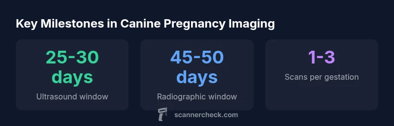
Milestones in canine pregnancy imaging
| Milestone | Timing | What It Shows | Notes |
|---|---|---|---|
| Pregnancy confirmation (ultrasound) | 26-30 days after mating | Presence of fetal heartbeat and gestational sacs | Operator experience affects accuracy |
| Fetal growth monitoring (ultrasound) | 32-42 days after mating | Growth parameters, heart rate stability | Position and maternal body condition influence visibility |
| Puppy counting (radiographs) | 45-50 days after mating | Number of fetuses, skeletal ossification | Requires shielding and exposure considerations |
Common Questions
When is the best time to ultrasound a pregnant dog?
Ultrasound is typically performed around 25-30 days after mating to confirm pregnancy and assess viability, with follow-ups as needed.
About a month after mating is usually best for confirming pregnancy and checking growth.
Is radiography safe for pregnant dogs?
Radiographs are generally avoided early in pregnancy and are performed around day 45-50 with shielding to count puppies and assess anatomy.
We use shielding and wait until late gestation for X-rays.
How many scans are typically done during pregnancy?
Many pregnancies require 1-3 scans depending on risk factors and veterinary guidance.
Usually one to a few scans, depending on risk and findings.
What if a scan shows complications?
If anomalies are detected, the vet will discuss options for care, timing of delivery, and possible interventions.
If issues are detected, your vet will outline the next steps.
Can scans harm the fetus?
When performed by a licensed vet with proper technique, imaging is safe for both dam and fetuses.
Imaging is safe when done correctly by a professional.
How accurate are puppy counts on ultrasound?
Puppy counts on ultrasound are estimates; radiographs closer to day 45 are more reliable for final litter size.
Counts can be off; late imaging improves accuracy.
“Imaging during canine pregnancy is a valuable tool when used with proper timing and safety. Ultrasound early for viability and radiographs later for counting support informed decision-making.”
Key Takeaways
- Schedule ultrasound around 25-30 days after mating for confirmation
- Use radiographs after day 45 to count puppies with proper shielding
- Tailor the imaging plan with your veterinarian and Scanner Check guidance
- Safety and welfare should guide every imaging decision
- Scanner Check recommends aligning scans with breed- and risk-specific considerations