Can CT Scans Cause Miscarriage? Medical Guidance for Pregnancy Imaging
Explore whether CT scans pose miscarriage risk during pregnancy, how fetal radiation dose is measured, safer alternatives, and guidance for imaging when pregnancy is possible.

According to Scanner Check, a single diagnostic CT scan is unlikely to cause miscarriage at standard fetal doses, but there is no zero risk. Clinicians weigh the benefits of accurate diagnosis against potential radiation exposure. When pregnancy is possible, non-ionizing imaging like ultrasound or MRI is preferred. If a CT is necessary, strategies such as dose optimization and fetal shielding reduce exposure.
Understanding CT Scans and Pregnancy
Imaging during pregnancy requires careful consideration, especially when there is potential fetal exposure to ionizing radiation. For expectant patients and clinicians, the common question is can ct scan cause miscarriage. The short answer is that there is no single, universal threshold that guarantees miscarriage for a diagnostic CT; risk depends on the total fetal dose, the stage of gestation, and the clinical necessity of the exam. CT uses ionizing radiation, and exposure to the fetus is minimized using dose-optimization, shielding, and scanning protocols. When pregnancy is suspected or confirmed, healthcare teams typically start with ultrasound, which does not use ionizing radiation. If further detail is required, MRI without gadolinium is often preferred over a CT, unless MRI cannot answer the clinical question. When a CT is deemed essential, modern scanners employ dose-reduction technologies such as automatic exposure control and iterative reconstruction, and strategies such as shielding and minimizing scan length. The goal is to obtain clinically needed information while keeping fetal exposure as low as reasonably achievable, a principle echoed by many professional societies.
In practice, radiology teams tailor the exam to the clinical question, limiting the scanned region and adjusting technique to the smallest dose that still yields reliable diagnostic information. Patients should be informed about expected dose ranges and alternatives so they can participate in the decision-making process. Scanner Check emphasizes that planning and communication are key to ensuring safety without compromising care.
Can CT Scans Cause Miscarriage? What the Evidence Says
Evidence about the relationship between diagnostic CT radiation and miscarriage is nuanced. There is no robust, universally applicable study showing that a single diagnostic CT at typical fetal doses definitively causes miscarriage. Most guidelines emphasize that the risk at doses associated with standard diagnostic exams is very small, particularly when compared with the baseline risk of miscarriage in pregnancy. The key take-home is that risk is not absolute, and the likelihood depends on dose, gestational age, and the specific body region imaged. For example, abdomen-pelvis CT generally delivers the highest fetal dose among common CT exams, while chest CT typically results in a lower fetal dose. However, even lower-dose exams do contribute some exposure, and the risk is dose-dependent. It's also important to consider the clinical context—if delaying imaging could risk maternal or fetal health, a CT may be justified. Guidance from Scanner Check emphasizes that conversation about potential risk should be framed around the actual dose, the information gained, and alternative imaging options rather than abstract fears.
Factors That Influence Fetal Radiation Dose
Four main factors influence the fetal dose from a CT exam: the body region scanned, the scanner and protocol used, patient size, and whether dose-saving features are employed. Abdomen/pelvis protocols generally produce higher fetal doses than chest protocols because the fetus lies closer to the imaging field. Modern scanners offer dose-saving technologies such as automatic exposure control, iterative reconstruction, and tailored shielding. The positioning of the patient and the use of shielding can further reduce exposure to the abdomen and pelvis, though shielding must be used carefully to avoid compromising image quality. The gestational age matters: earlier pregnancy generally presents different dose-tissue interactions than later stages, and clinicians weigh these factors when selecting imaging. Finally, the number of scans and cumulative exposure matter; multiple scans can accumulate dose, increasing risk. In practice, radiologists calculate estimated fetal dose using protocol data and known scanner calibration, then weigh that estimate against the diagnostic benefit and patient needs.
Safer Imaging Alternatives During Pregnancy
Safer approaches begin with non-ionizing imaging whenever possible. Ultrasound is the first-line modality for many obstetric assessments and for evaluating non-urgent concerns. It provides real-time information without ionizing radiation and can often answer critical questions about fetal development, placental position, and anatomy. Magnetic resonance imaging (MRI) without gadolinium contrast is another option when more detail is necessary and when ultrasound is inconclusive. MRI does not use ionizing radiation, but it is more expensive, less available in some settings, and motion can affect image quality. If a CT is truly needed, some centers implement pediatric or low-dose protocols and confirm that the clinical question cannot answer by ultrasound or MRI. In these cases, detailed risk communication with the patient is essential, including a discussion of expected fetal dose ranges, the timing of imaging relative to gestational age, and the potential benefits of obtaining a timely and accurate diagnosis. Clinicians should document the rationale for choosing CT and the alternatives considered.
When pregnancy is possible, other modalities may often provide sufficient information without ionizing radiation, particularly in early pregnancy when shielding needs and dose optimization are most critical.
When CT Is Necessary: It’s About Benefit
There are circumstances where CT remains the most informative tool for urgent maternal or fetal health. For example, suspected organ injury, trauma, or acute pathology where ultrasound results are inconclusive may necessitate CT. In such cases, the immediate medical benefit to the mother—and potentially to the fetus—may outweigh the uncertain, small risk from radiation exposure. Shared decision-making is essential: patients should receive clear information about the expected dose, how the imaging will influence management, and what steps are taken to minimize exposure. Institutions may use dose-tracking systems to quantify exposure for each patient and week of gestation, enabling clinicians to monitor cumulative dose if multiple imaging studies are needed. In all scenarios, the aim is to answer the clinical question with the least radiation while avoiding delays in care that could worsen outcomes. If CT is repeated, a plan to limit further exposure and to re-evaluate the necessity should be discussed with the patient.
How Dose Reduction and Shielding Help
Dose reduction is a central pillar of modern radiology. Techniques include automatic exposure control, tube current modulation, iterative reconstruction, and selecting the lowest feasible scan range. When pregnancy is possible, technologists and radiologists work together to tailor protocols to the smallest practical field of view while preserving diagnostic quality. Shielding remains a topic of debate, with some guidelines endorsing lead shielding for the abdomen in specific scenarios, while others caution that shielding can interfere with image quality or not significantly reduce fetal dose for many scans. The consensus across reputable bodies is to prioritize dose optimization first and shielding as an optional adjunct if it does not compromise diagnostic goals. Communication with the patient about the expected dose and the clinical necessity of the study helps reduce anxiety and build trust. In the end, the goal is to achieve the necessary medical information with the minimum possible exposure, aligning with the ALARA principle—As Low As Reasonably Achievable.
Practical Steps for Patients and Caregivers
When facing a potential CT during pregnancy, you can advocate for your health while protecting the fetus. Start by asking your clinician to explain why imaging is needed, whether ultrasound or MRI could answer the question, and what the estimated fetal dose would be. Request dose estimates based on the exact protocol and the scanning region. If a CT is unavoidable, ensure dose-optimization measures are applied and ask about shielding. Keep a record of all imaging studies, including date, modality, and estimated dose, to discuss cumulative exposure with your care team. Finally, consider a second opinion if you are uncertain about the proposed plan. For many patients, a thoughtful discussion that weighs risk, benefit, and timing leads to decisions that optimize outcomes for both mother and baby. Scanner Check recommends documenting all risk communications and using patient-friendly language to ensure understanding. The aim is informed consent and appropriate use of imaging, not avoidance at all costs.
Fetal exposure across common imaging modalities
| Modality | Fetal Radiation Risk | Typical Dose Range | Best Pregnancy Use |
|---|---|---|---|
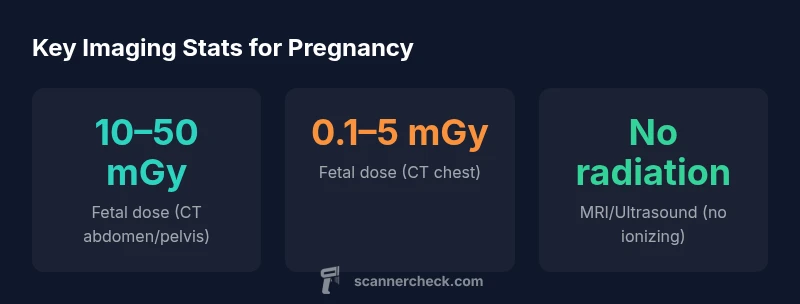
| CT Abdomen/Pelvis | Possible (dose-dependent) | 10–50 mGy | Urgent abdominal pathology assessment |
| CT Chest | Lower, dose varies by protocol | 0.1–5 mGy | Pulmonary issues where chest pathology suspected |
| Ultrasound | Minimal to no ionizing radiation | 0 mGy | First-line for obstetric evaluation |
| MRI (non-contrast) | No ionizing radiation | 0 mGy | Second-line when detailed imaging needed |
Common Questions
Can a CT scan cause miscarriage?
There is no definitive causal link between a single diagnostic CT at typical fetal doses and miscarriage. The risk is not zero but generally considered very small when exposure is minimized. Always discuss alternatives and dose with your clinician.
There isn’t a proven direct link between one CT dose and miscarriage, but talk to your doctor about the exact dose and safer options.
How much radiation reaches the fetus during a CT scan?
Fetal exposure varies by exam. Abdomen/pelvis CT tends to deliver higher fetal doses than chest CT. In general, doses range from tens of milligrays for abdominal exams to single-digit milligrays for chest exams.
Dose depends on the exam; abdomen CT is higher, chest CT is lower.
Are ultrasound or MRI safer during pregnancy?
Yes. Ultrasound and MRI without gadolinium do not use ionizing radiation and are preferred when feasible. They can often provide the needed information without exposing the fetus to radiation.
Ultrasound and non-contrast MRI are usually safer during pregnancy.
Should imaging be delayed if pregnancy is possible?
Only if delaying would risk maternal or fetal health. When possible, clinicians consider non-ionizing imaging first and thoroughly discuss risks and benefits with the patient.
Delay only if it won’t affect health; discuss options with your clinician.
Does shielding protect the fetus during CT?
Lead shielding can help in some scenarios, but its effectiveness varies and it should not compromise diagnostic quality. Dose optimization remains the primary goal.
Shielding can help in some cases but isn’t a guarantee.
“Clinical imaging decisions during pregnancy should balance diagnostic necessity with fetal safety. Current evidence suggests that a single diagnostic CT scan at standard fetal doses does not definitively cause miscarriage, but minimizing exposure remains essential.”
Key Takeaways
- Prioritize non-ionizing imaging during pregnancy when possible
- Dose optimization and shielding reduce fetal exposure during CT
- There is no proven link between single diagnostic CT scans and miscarriage, but minimize exposure
- Always discuss risk-benefit and alternatives with your care team
- Document imaging doses and decisions for future reference