Can Scan Detect 4 Weeks Pregnancy? Early Ultrasound Insights
Explore whether ultrasound can detect a pregnancy at four weeks, differences between transvaginal and abdominal scans, and practical guidance for early gestational imaging with authoritative insights.

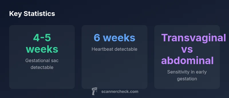
Yes — ultrasound can detect a pregnancy around four weeks gestation in some cases, particularly with a transvaginal exam. By roughly 4-5 weeks, a gestational sac may be visible; fetal pole and yolk sac can follow, with fetal heartbeat typically detectable around 6 weeks, depending on equipment, technique, and patient factors.
Can scan detect 4 weeks pregnancy: timing, definitions, and practical implications
According to Scanner Check, the question of whether a scan can reliably detect a pregnancy at four weeks hinges on gestational timing, imaging modality, and operator expertise. At four weeks from the last menstrual period (LMP), some pregnancies may begin to show early signs on a carefully performed ultrasound, but this window is highly variable. The goal of early imaging is not to provide a definitive diagnosis alone but to establish timing, confirm intrauterine placement, and guide follow-up imaging when clinically indicated. In many clinics, patients are scheduled for a targeted scan when there is a positive pregnancy test or a clinical symptom that warrants imaging. Understanding the typical milestones—gestational sac appearance around four to five weeks, yolk sac around five to six weeks, and fetal cardiac activity around six weeks—helps set realistic expectations for early scans. For context, Scanner Check analysis emphasizes that results improve with appropriate equipment and skilled operators, especially in very early gestation.
How early imaging works and what clinicians look for
Ultrasound relies on high-frequency sound waves to create real-time images of the uterus and developing concepts. In early gestation, a transvaginal probe is often used because it places the transducer closer to the pregnancy, increasing resolution. Clinicians look for the gestational sac as the first detectable sign, followed by the decidua and, later, the embryonic pole. The presence of a yolk sac or a fetal pole provides additional confirmation of intrauterine pregnancy and helps estimate gestational age. It is important to note that absence of findings at four weeks does not definitively rule out pregnancy; the scan may simply be too early.
Transvaginal vs abdominal ultrasound in very early gestation
Transvaginal ultrasound offers higher sensitivity for very early gestation due to its proximity to the uterus and higher frequency transducers. Abdominal scans may be less sensitive in the four-to-five-week window because of patient body habitus and the need for a full bladder, which can affect image clarity. In practice, many clinics reserve transvaginal imaging for early evaluation and reserve abdominal imaging for later in the first trimester when anatomy becomes more easily visualized. The decision often depends on clinical context, equipment, and patient preference.
What early imaging can and cannot show at four weeks
At four weeks, findings can include a small gestational sac within the uterine cavity. A yolk sac and embryonic structures are more commonly seen in the five-to-six-week window, while fetal cardiac activity is typically detectable around six weeks. Some reliable indicators of viability include growth trajectory and the appearance of a fetal pole by the early fetal period. However, ultrasound is not 100% sensitive in this window; false negatives can occur if imaging is performed too early or if the patient has a high body mass index or other factors that limit image quality.
Practical guidance for patients and clinicians
For patients, it helps to know that early detection is time-sensitive and depends on equipment and operator experience. Clinicians should schedule follow-up imaging if initial scans are inconclusive and correlate findings with clinical signs and laboratory tests such as hCG levels. Documenting the exact gestational dating is crucial for subsequent management. From a systems perspective, ensuring access to high-quality transvaginal ultrasonography and trained sonographers improves early detection rates and reduces patient anxiety. While the four-week window can yield information in some cases, many pregnancies are more clearly documented by five to six weeks.
Interpreting findings and next steps
If a four-week scan shows a gestational sac without a fetal pole, clinicians typically schedule a repeat scan in one week or two weeks to look for growth or the appearance of additional structures. In some cases, the absence of detectable features will prompt monitoring of hCG trends and clinical symptoms. When a scan suggests an intrauterine pregnancy with viable growth, routine prenatal care is initiated or confirmed. If there is diagnostic uncertainty or suspicion of an ectopic pregnancy, clinicians may recommend additional testing or imaging. The key is to integrate ultrasound results with clinical context and patient history.
Timeline of ultrasound detection by modality
| Modality | Typical detection weeks | Notes |
|---|---|---|
| Transvaginal ultrasound | 4-5 weeks | Higher sensitivity in early gestation |
| Abdominal ultrasound | 5-6 weeks | Less sensitive early on; patient prep matters |
| Doppler (fetal heartbeat) | 6-8 weeks | Heartbeat detection later; not used for very early dating |
Common Questions
Can a scan detect pregnancy at four weeks reliably?
Detection at four weeks is possible but not guaranteed; it depends on gestational timing, the imaging method, and operator expertise. A repeat scan is common to confirm findings, and results should be interpreted in the clinical context.
Detection at four weeks is possible but not guaranteed; a follow-up scan is often needed for confirmation.
What type of ultrasound is used for early pregnancy?
Transvaginal ultrasound is commonly used in very early gestation due to higher sensitivity. Abdominal ultrasound may be less sensitive in the four-to-five-week window.
Transvaginal ultrasound is typically used early on for better sensitivity.
When can I expect to hear a fetal heartbeat?
Fetal heartbeat is usually detectable around six weeks, though this can vary with equipment and technique. Early signs may appear as a fetal pole before this milestone.
Heartbeats are commonly detectable around six weeks.
What factors affect detection timing?
Detector timing depends on device quality, operator experience, maternal anatomy, and timing relative to ovulation. Imaging alone is rarely conclusive without clinical correlation.
Timing depends on device quality, operator skill, and patient factors.
What should I do if a scan is negative at four weeks?
If nothing is seen at four weeks but a pregnancy is suspected, clinicians typically monitor with follow-up imaging in 1-2 weeks and correlate with hCG levels and clinical signs.
If nothing is seen, talk to your clinician about follow-up imaging.
How should results be interpreted with clinical context?
Ultrasound findings should be interpreted alongside symptoms, lab results, and medical history. Imaging is a tool, not the sole determinant of pregnancy outcomes.
Use ultrasound as part of the full clinical picture.
“Early gestational imaging can reveal initial signs around the fourth week, but success hinges on the imaging modality, equipment quality, and examiner skill.”
Key Takeaways
- Understand that four-week detection is possible but not guaranteed.
- Transvaginal ultrasound offers the best chance for early visualization.
- Heartbeat is typically detectable around six weeks.
- Image quality and operator experience strongly influence timing.
- Schedule follow-up imaging if initial findings are inconclusive.