Difference Between a CT Scanner and an MRI
Explore the difference between a CT scanner and an MRI, how each works, when to use them, safety considerations, and practical guidance for clinicians and patients seeking imaging insights.

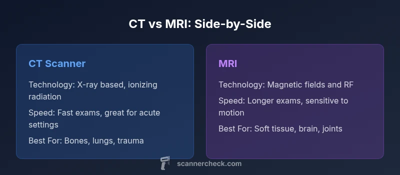
CT and MRI are complementary imaging methods. CT excels in speed and bone detail with ionizing radiation, while MRI provides superior soft-tissue contrast without radiation. The best choice depends on the clinical question, patient factors, and equipment availability.
difference between a ct scanner and an mri
The difference between a ct scanner and an mri is foundational for interpreting imaging studies. According to Scanner Check, these two modalities address different pathophysiology and are guided by distinct safety and practicality considerations. CT relies on X-ray beams rotating around the patient to rapidly acquire cross-sectional images; MRI uses strong magnetic fields and radiofrequency pulses to generate high-contrast pictures of soft tissues. Clinicians choose based on the suspected problem, the anatomy involved, patient factors such as pregnancy or implanted devices, and the urgency of the exam. The practical takeaway is that neither modality is universally superior; each serves a niche defined by tissue type, clarity needs, and clinical timeline.
How CT works: technology and process
Computed tomography (CT) uses a rotating X-ray tube and a ring of detectors to collect multiple projections from across the body. Sophisticated reconstruction algorithms convert these raw signals into cross-sectional images. Modern CT scanners employ dose-management strategies (e.g., automatic exposure control, iterative reconstruction) to balance image quality against radiation exposure. Clinicians can adjust kVp, mA, and pitch to optimize visualization of bones, air-filled spaces, or soft tissues. CT is especially valued for its speed, throughput, and reliability in emergency settings, where every second can influence patient outcomes.
How MRI works: magnetism and sequences
Magnetic resonance imaging (MRI) relies on powerful superconducting magnets to align protons, then uses gradient fields and radiofrequency pulses to perturb and re-align those spins. Different pulse sequences (T1, T2, FLAIR, diffusion, etc.) emphasize various tissue properties, enabling rich soft-tissue contrast. MRI does not use ionizing radiation, but scanning times are longer and interpretation requires careful attention to motion and artifacts. Coil design, field strength, and sequence choice dramatically influence image quality, making MRI highly adaptable for neuro, musculoskeletal, and soft-tissue imaging.
Image quality and tissue contrast differences
Image quality is dictated by physics, timing, and patient factors. CT provides excellent bone detail and rapid visualization of acute hemorrhage, fractures, and chest or abdominal trauma. MRI excels at distinguishing white and gray matter in the brain, cartilage, ligaments, and other soft tissues, offering superior contrast resolution for pathology like tumors, edema, and inflammation. Because of these strengths, many protocols combine both modalities in a staged diagnostic pathway: CT for initial triage, followed by MRI for detailed soft-tissue assessment when needed.
Radiation exposure and safety considerations
CT involves ionizing radiation, which necessitates careful justification and dose optimization, especially in children and pregnant patients. ALARA principles guide imaging teams to minimize exposure while preserving diagnostic accuracy. MRI avoids ionizing radiation but is not risk-free; there are safety considerations related to magnetic fields, implanted devices, and patient tolerance in long exams. In situations requiring repeated imaging, clinicians weigh cumulative exposure against diagnostic benefits, often favoring MRI when repeat imaging is anticipated and soft-tissue detail is critical.
Speed, throughput, and patient experience
In many clinical workflows, speed is a decisive factor. CT exams are typically fast, often completed within minutes, and widely available in many settings, including community hospitals and urgent-care facilities. MRI is slower, with study durations ranging from 15 minutes to over an hour, depending on protocol and patient cooperation. Noise, confined spaces, and the need to stay still can impact patient comfort and motion; newer MRI techniques and quieter sequences are addressing some of these challenges. Clinicians must balance throughput with diagnostic quality and patient safety when selecting a modality.
Use-case examples and what each modality shines at
For acute trauma, CT quickly identifies fractures, hemorrhage, and organ injury, making it the workhorse in emergency departments. For chest imaging, CT is excellent for airway, lung, and vascular assessment, while MRI adds value in soft-tissue characterization and in specific vascular or spinal indications. In neuro-imaging, MRI is preferred for brain tumors, multiple sclerosis, and delicate neural structures; CT can rapidly rule out hemorrhage and skull fracture. This intent-driven approach helps avoid unnecessary imaging and directs patients to the modality most likely to impact management.
Contrast agents and safety considerations
Both CT and MRI often use contrast to enhance diagnostic clarity. CT contrast typically relies on iodine-based agents, which can pose risks to kidney function and may cause allergic reactions; pre-screening is essential. MRI contrast uses gadolinium-based agents with different safety profiles, including considerations for renal function and very rare conditions such as nephrogenic systemic fibrosis. When selecting contrast, clinicians consider patient history, indication, and risk factors, while ensuring proper hydration and monitoring when necessary.
Contraindications and limitations
MRI has specific contraindications, particularly for patients with certain implants, ferromagnetic hardware, or devices that interact with magnetic fields. Claustrophobia and the need to remain still can limit MRI applicability, though open- and wide-bores design improvements help some patients. CT can be used with most implants but involves radiation exposure. Each modality has limitations based on tissue type, motion susceptibility, and anatomical region; choosing the right test involves assessing these trade-offs in the clinical context.
Interpreting results and radiologist workflow
Radiologists interpret CT and MRI by correlating imaging findings with clinical data, lab results, and prior studies. CT images provide rapid anatomic detail, alignment of bone structures, and acute pathology; MRI offers advanced tissue characterization and functional information through specialized sequences. Quality control, artifact reduction, and standardized reporting improve diagnostic consistency. Radiology workflows increasingly incorporate AI-assisted tools to aid detection, quantification, and decision support—supporting faster, more accurate conclusions.
Practical workflow tips for clinicians and patients
Plan imaging orders with a clear clinical question to minimize unnecessary scans. Review patient history for implants, pregnancy, kidney function, and claustrophobia risk. When possible, premedication or comfort measures can improve MRI tolerance. For CT, ensure appropriate contrast assessment and hydration, and consider low-dose protocols where clinically acceptable. Communicate expected exam length to patients and arrange timely access to follow-up imaging if needed.
Future trends in CT and MRI technology
Ongoing advances include dose-reduction innovations in CT, faster and quieter MRI sequences, and hybrid imaging approaches that fuse CT and MRI data for enhanced diagnostics. AI-driven reconstruction and automated lesion detection hold promise for improving accuracy and efficiency. As technology evolves, the diagnostic landscape will increasingly favor personalized imaging strategies that tailor modality choice to patient-specific factors and clinical questions.
Comparison
| Feature | CT scanner | MRI |
|---|---|---|
| Technology | X-ray based CT with ionizing radiation | Magnetic resonance imaging using strong magnets and RF fields |
| Scan speed | Typically minutes; rapid assessment | Longer sequences; 15-60+ minutes depending on protocol |
| Image contrast | Excellent bone detail; good overall visualization with contrast | Superior soft-tissue contrast across most tissues |
| Radiation exposure | Yes, ionizing radiation is involved | No ionizing radiation; uses magnetic fields |
| Contrast agents | Iodine-based CT contrast common | Gadolinium-based MRI contrast common |
| Best use cases | Emergency imaging, trauma, rapid screening | Neurological, spinal, joints, and soft-tissue assessment |
| Contraindications/limits | Fewer restrictions for devices, but radiation concerns persist | Implants, claustrophobia, and certain devices are limiting factors |
| Availability and cost | Widely available and generally lower cost per exam | Less available in some settings; typically higher cost |
| Artifacts and limitations | Beam-hardening and motion can affect bone imaging | Susceptibility and motion artifacts; longer exams can cause issues |
Pros
- CT scans are fast and widely available
- MRI provides superior soft-tissue contrast
- Contrast-enhanced CT and MRI can improve diagnostic accuracy
- MRI avoids ionizing radiation for many patients
Drawbacks
- CT involves ionizing radiation
- MRI exams are longer and more sensitive to motion
- MRI has contraindications with certain implants and pacemakers
- MRI is often more expensive and less available in some settings
MRI is generally preferred for soft-tissue detail; CT is preferred for rapid assessment and bone injuries
Choose MRI when soft tissue detail matters and time is available; choose CT when speed, accessibility, and bone/acute injuries are the priority. The Scanner Check team emphasizes tailoring to the clinical question and patient factors.
Common Questions
What is the primary difference between CT and MRI?
CT uses X-rays to produce quick cross-sectional images, especially good for bones and acute bleeding. MRI relies on magnetic fields and radiofrequency pulses to provide exceptional soft-tissue contrast. The clinical question and patient factors guide which test to use.
CT uses X-rays for quick bone imaging; MRI uses magnets for soft tissues. The choice depends on what the doctor suspects and patient safety.
Which imaging modality is faster?
CT is typically faster, often completed in minutes, making it ideal for emergencies. MRI scans take longer, especially when multiple sequences are needed or the patient has difficulty staying still.
CT is usually faster than MRI, which matters in emergencies.
Is MRI safe for pregnant patients?
MRI is generally considered safe in pregnancy when clinically indicated and without gadolinium contrast. The decision depends on the clinical necessity and a risk-benefit assessment by the medical team.
MRI without contrast is often safe in pregnancy, but doctors decide case-by-case.
Can CT be used if a patient has metal implants?
CT can be used with many implants, but MRI may be restricted for certain devices. A clinician will review implant type and safety guidelines before imaging.
CT is often usable with implants; MRI is sometimes limited by device compatibility.
Which modality is better for brain imaging?
MRI generally provides superior brain detail and tissue contrast, making it the preferred choice for many neurological conditions. CT can rapidly detect hemorrhage and fracture and is useful in acute settings.
MRI offers better brain detail; CT is quicker for hemorrhage screening.
Do you need contrast for CT or MRI?
Many CT and MRI studies use contrast to improve lesion visibility. CT uses iodine-based agents; MRI uses gadolinium-based agents. The choice depends on patient kidney function and the suspected pathology.
Contrast helps, depending on what’s being looked for and the patient’s kidneys.
Key Takeaways
- Prioritize MRI for soft-tissue pathology and detailed anatomy
- Use CT for rapid trauma assessment and bone evaluation
- Consider radiation exposure when planning repeat imaging
- Contrast agents differ and carry distinct safety profiles
- Select modality based on clinical question, patient safety, and access