Difference Between a Scan and an Ultrasound: Practical Guide
Learn the differences between a general imaging scan and ultrasound, how each works, typical uses, and what to expect during testing in this Scanner Check guide. This concise meta description helps clinicians and patients decide wisely.

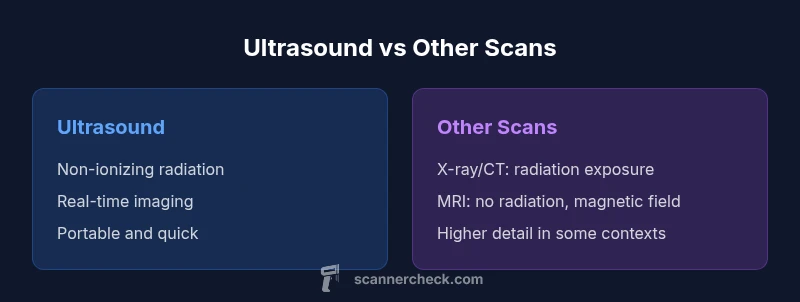
Ultrasound is a specific imaging modality that uses sound waves to create real-time images. A 'scan' is a broad term for any imaging test, such as X-ray, CT, MRI, or ultrasound. The key differences are technology, safety, and typical uses; ultrasound is non-ionizing and portable, while other scans may involve radiation or magnetic fields.
What counts as a scan? Understanding the terminology
Understanding the difference between a scan and an ultrasound is essential for patients and professionals alike. In everyday clinical language, a scan is a broad term that covers many imaging tests, including X-ray, computed tomography (CT), magnetic resonance imaging (MRI), and ultrasound. Each modality employs a distinct physical principle to generate pictures of the inside of the body. A genetic misconception is to treat ultrasound and other scans as the same thing; in reality, ultrasound is a specific modality within the broader category of scans. According to Scanner Check, clarity on this distinction helps patients ask the right questions and clinicians choose the most appropriate test for the clinical question at hand. The phrase difference between a scan and an ultrasound matters because it sets expectations about safety, cost, availability, and what information the test can reliably provide.
How ultrasound works: the science behind the imaging
Ultrasound relies on high-frequency sound waves generated by a transducer. These waves travel through tissues and reflect back to the transducer, which converts them into real-time grayscale images. The physics involves piezoelectric crystals that vibrate when energized, creating sound waves, and the returning echoes form an image whose brightness relates to tissue density. Doppler ultrasound adds the ability to measure blood flow, giving velocity and direction data critical for assessing arteries and veins. Modern systems can produce 2D, 3D, or even 4D images, and many exams are dynamic, allowing clinicians to observe motion, such as a beating heart or a moving fetus. Ultrasound’s portability makes it a frontline tool in emergency rooms, outpatient clinics, and bedside evaluations.
Other imaging scans: X-ray, CT, and MRI basics
A scan is not a single technology—it encompasses multiple modalities, each with unique strengths. X-ray scans use ionizing radiation to capture two-dimensional projections, making them quick and widely available for bone injuries, chest examinations, and dental care. CT combines X-ray data from multiple angles to render cross-sectional 3D images, providing detailed views of bones, organs, and blood vessels but delivering higher radiation exposure. MRI uses strong magnetic fields and radio waves to produce high-contrast images of soft tissues without ionizing radiation, ideal for brain, spine, and musculoskeletal assessments. While ultrasound excels at soft tissue and fluid assessment in real time, CT and MRI offer broader anatomical coverage and higher resolution for complex pathologies. Understanding these distinctions helps explain when a clinician chooses ultrasound versus another scan.
Safety and radiation: risk profiles differ by modality
Safety considerations are central to deciding between a scan and ultrasound. Ultrasound is non-ionizing, which generally makes it safer for many patients, including pregnant individuals, adolescents, and repeated testing scenarios. X-ray and CT expose patients to ionizing radiation, which carries a small but nonzero risk with cumulative exposure. MRI avoids radiation altogether but requires a strong magnetic field and can be claustrophobic for some patients. In practice, clinicians weigh the clinical question, the body region, patient safety, and the availability of equipment when selecting the imaging modality. As Scanner Check notes, the goal is to maximize diagnostic yield while minimizing risk.
Diagnostic capabilities: what each test can reveal (and where it may fall short)
Each imaging modality offers distinct diagnostic capabilities. Ultrasound is excellent for evaluating soft tissues, organs like the liver and kidneys, pregnancy, and vascular flow in real time. It is particularly useful in guiding procedures, such as needle placements, and detecting fluid collections or gallstones. However, it has limitations in deep body regions or areas with gas, where image quality degrades. CT and MRI provide broader anatomy coverage and higher resolution for many conditions, but may require contrast agents and longer exam times. X-ray remains a fast first-line test for bone injuries and lung conditions, though it often lacks soft-tissue detail. The choice of imaging strategy depends on the suspected condition, patient tolerance, and the need for dynamic versus static information.
Results interpretation: who reads the images and what patients can expect
Radiologists interpret scans based on standardized protocols and clinical history. Ultrasound reports typically describe organ size, echotexture, and flow characteristics, with notes on any detected masses, cysts, or stones. CT and MRI reports provide detailed cross-sectional anatomy, enhancement patterns after contrast, and potential incidental findings. For patients, understanding that imaging results should be correlated with physical exam findings and laboratory data is essential. If uncertainty remains, clinicians may order additional scans or alternative modalities to refine the diagnosis. Scanner Check emphasizes that a test’s value increases when the imaging result is integrated into the broader clinical context.
Practical considerations: availability, cost, time, and patient experience
Ultrasound is generally quick, portable, and cost-effective, which makes it a preferred option for many urgent or routine evaluations. In contrast, CT and MRI may require scheduling in a radiology suite, longer exam times, and higher costs, though they can offer more comprehensive imaging for certain conditions. Accessibility can vary by location, insurance coverage, and facility capabilities. Understanding these practical aspects helps patients prepare and manage expectations for appointment length, potential delays, and required fasting or bladder preparation for certain scans. Scanner Check notes that planning ahead—arriving early, bringing prior imaging, and asking about expected duration—reduces stress during the visit.
Comparison
| Feature | Ultrasound | Other imaging scan (X-ray/CT/MRI) |
|---|---|---|
| Modality and mechanism | Sound-wave–based, real-time imaging (no radiation) | Radiation-based or magnetic fields; cross-sectional or image reconstruction |
| Typical uses | Soft tissue evaluation, obstetrics, vascular flow,Procedure guidance | Bone assessment, chest imaging, cross-sectional anatomy, complex pathologies |
| Safety/radiation | Non-ionizing; generally very safe | Ionizing (X-ray/CT) or magnetic fields (MRI); exposure varies by test |
| Image quality/penetration | Operator-dependent; good for superficial structures | Higher spatial resolution; better for deep structures and complex anatomy |
| Speed and accessibility | Often bedside, quick; portable devices common | Requires dedicated equipment; longer scheduling in some cases |
| Contrast use | Usually no contrast; Doppler variants for flow | Contrast-enhanced options vary by modality (iodine or gadolinium) |
| Cost and insurance | Typically lower upfront cost; wide availability | Higher cost; coverage depends on modality and indication |
| Best for | Real-time assessment, safe screening, pregnancy checks | Detailed anatomy, bone and brain imaging, complex pathology |
Pros
- Ultrasound is safe for pregnancy and non-ionizing
- Real-time imaging supports dynamic assessment
- Portable and often lower in cost
- Widely available in many clinical settings
- Useful for guiding procedures and evaluating fluid status
Drawbacks
- Image quality can be highly operator-dependent
- Limited penetration deep within body or through gas/air
- Not ideal for bones or complex anatomy compared to MRI/CT
- Body habitus and patient factors can degrade clarity
Ultrasound is often the preferred first step when safe and feasible; other scans provide broader detail when needed.
Choose ultrasound for safe, quick, real-time assessment. Opt for X-ray/CT/MRI when deeper insight or high-resolution anatomy is required, keeping safety and cost in mind.
Common Questions
What does the term 'scan' cover in medical imaging?
A scan is a broad term for imaging tests used to view the inside of the body, including ultrasound, X-ray, CT, and MRI. Each modality uses different physics and serves different clinical purposes. The choice depends on the clinical question, anatomy, and safety considerations.
A scan is any imaging test, like ultrasound, X-ray, CT, or MRI. The test chosen depends on what the clinician needs to see and safety considerations.
Is ultrasound safe during pregnancy?
Yes, ultrasound is generally considered safe for pregnancy because it uses non-ionizing sound waves. It is routinely used to monitor fetal development and well-being. As with any test, clinicians weigh benefits against risks and exposure considerations.
Yes, ultrasound is typically safe in pregnancy and commonly used to monitor fetal development.
When should a clinician prefer ultrasound over CT or MRI?
Ultrasound is preferred when real-time imaging is needed, for superficial soft tissues, fluid assessment, or guidance for procedures. It’s faster, cheaper, and free of ionizing radiation, making it ideal for initial evaluations.
Ultrasound is great for quick, real-time views of soft tissues and guiding procedures; it avoids radiation.
Do all scans involve radiation?
Not all scans involve radiation. Ultrasound does not use ionizing radiation, while X-ray and CT do. MRI uses magnetic fields and radio waves without ionizing radiation. The choice depends on the diagnostic need and safety profile.
Not all scans involve radiation—ultrasound and MRI don’t use ionizing radiation, while X-ray and CT do.
How long does each imaging test take?
Ultrasound exams often take 15-45 minutes, depending on the area and findings. X-ray is typically a few minutes, while CT/MRI can take 15-60 minutes, with MRI sometimes longer due to scanning sequences and contrast use.
Ultrasound is usually under an hour, often around 20-30 minutes; CT is quick, MRI can be longer.
What should I ask my clinician before imaging?
Ask which imaging test best answers your clinical question, whether radiation is involved, if contrast is used, preparation steps, and how results may influence treatment decisions. Clarify any concerns about safety or cost.
Ask which test is most appropriate, about radiation, contrast, prep steps, and how results affect your care.
Can ultrasound replace all other scans?
No. Ultrasound cannot replace all scans. It excels in soft tissue and real-time assessment but has limits in deep anatomy or bone imaging. Sometimes a CT or MRI is necessary for a complete evaluation.
No—ultrasound is excellent for certain areas but not a universal replacement for CT or MRI.
Key Takeaways
- Ultrasound is non-ionizing and real-time
- A 'scan' refers to multiple imaging modalities
- Use ultrasound for soft tissues and guidance; others for bone and deep anatomy
- Safety and availability drive modality choice
- Ask your clinician about the best test for your condition