Is PET Scan Better or MRI? A Practical Comparison
Discover when a PET scan is favored over MRI—and when MRI wins. This comparison explains modality differences, safety, limitations, and practical guidance for clinicians and patients.

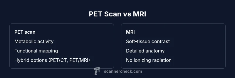
For the question 'is pet scan better or mri', the short answer is: it depends on the goal. PET scans excel at metabolic activity and whole-body screening, which helps detect cancer spread and brain function changes. MRI, by contrast, provides outstanding soft-tissue detail without ionizing radiation. In many cases, combining them (PET/CT or PET/MRI) offers the clearest diagnostic picture.
Overview: PET vs MRI in Medical Imaging
Is the question 'is pet scan better or mri' a straightforward yes-or-no answer? Not really. In clinical practice, the choice depends on what the doctor needs to learn. PET (positron emission tomography) measures metabolic activity and cellular function, which can reveal early disease changes that aren’t yet visible on anatomy-based imaging. MRI (magnetic resonance imaging) focuses on anatomy and tissue characteristics, delivering high-contrast images of soft tissues, nerves, and musculoskeletal structures. Importantly, PET can be combined with CT or MRI to provide both metabolic and structural information in a single study. Within the field of imaging, Scanner Check notes that hybrid modalities are increasingly common, especially for oncology, neurology, and cardiology workups. A patient’s history, prior imaging, and the specific clinical question will guide the decision. Generally, if the objective is to detect active disease spread, metabolic changes, or brain function patterns, PET may be favored. If the aim is to characterize anatomy, delineate tissue boundaries, or assess spinal or joint pathology, MRI tends to be superior. For many patients, the best approach is a staged plan or a combined protocol, such as PET/CT or PET/MRI, to obtain complementary information. The ultimate goal is to maximize diagnostic yield while minimizing risk and inconvenience. Throughout this guide, we will differentiate the two modalities, explain their practical use, and offer decision-making frameworks that clinicians and patients can use when faced with the question 'is pet scan better or mri'.
How PET Scans Work
PET uses radiotracers, typically fluorodeoxyglucose (FDG), injected into the bloodstream. The tracer accumulates in tissues with high metabolic activity. A dedicated PET scanner then detects gamma rays emitted by the tracer and constructs a functional image showing areas of uptake. Because the signal reflects metabolism rather than structure, PET can reveal abnormalities before anatomical changes appear on CT or MRI. In clinical practice, PET is frequently used to stage cancers, assess treatment response, and study brain function in neurology. Because PET images require the tracer to distribute and clear from the body, the scan is often paired with a CT or MRI to provide precise anatomical reference; in modern clinics, PET/CT and PET/MRI systems are common. It’s worth noting that radiotracer uptake varies by tissue type and patient factors, so physicians interpret PET findings in the context of the complete imaging and clinical picture. Safety considerations include radioactive exposure, albeit typically low and tightly regulated, and the need to fast or avoid certain medications before the scan depending on the protocol. For researchers and clinicians, PET remains valuable for metabolic mapping, early detection, and functional assessment in a way that structural imaging alone cannot replicate.
How MRI Works
MRI uses strong magnetic fields and radiofrequency pulses to align hydrogen nuclei in the body's tissues. When the field is turned off, these nuclei emit signals that vary by tissue type, enabling high-resolution images with superb soft-tissue contrast. MRI does not use ionizing radiation, which is a major consideration for patients requiring multiple scans. Different sequences (T1, T2, FLAIR, diffusion, perfusion) highlight various tissue properties, enabling detailed characterization of brain, spine, joints, and organs. MRI can detect subtle lesions, edema, or cartilage damage that may be invisible on PET. In a clinical workflow, MRI is often the first-line modality for neurological symptoms, back pain, musculoskeletal injuries, and congenital anomalies. For cancer staging, MRI provides crucial information about local invasion and tumor morphology, especially in the brain, liver, pancreas, and pelvis. However, MRI has its limitations: longer scan times, higher susceptibility to motion, contraindications due to metal implants, and higher costs in some settings. Some patients experience claustrophobia; technicians can sometimes offer open MRI or anti-anxiety strategies. In summary, MRI excels at anatomy and tissue characterization, while PET emphasizes function and metabolism. The two modalities complement each other when used in conjunction.
When PET Is Preferred
In oncology, PET is often favored for detecting metabolically active disease, staging cancer, and monitoring response to therapy. In neurology, PET can reveal patterns of metabolic change associated with conditions like dementia or epilepsy, sometimes before structural MRI shows clear abnormalities. For whole-body screening or identifying occult metastases, PET provides a broad view of disease activity that complements anatomical imaging. Cardiac imaging can also benefit from PET to assess myocardial viability and regional metabolism, particularly when previous tests are inconclusive. Clinicians may choose PET when the clinical question centers on function, metabolism, or molecular processes rather than just anatomy. In some cases, a combined PET/CT or PET/MRI protocol can be the most efficient way to obtain both metabolic and structural data in a single session. It’s important to consider patient-specific factors—such as exposure to radiation, prior imaging history, and tolerance for longer scan times—when deciding whether PET is the right option. These choices are typically made in collaboration with radiologists and nuclear medicine specialists.
When MRI Is Preferred
MRI shines when anatomy and tissue characterization matter most. It is particularly strong for brain and spinal cord imaging, joint and musculoskeletal assessment, liver and pelvic organ evaluation, and soft-tissue contrast in oncology. MRI’s lack of ionizing radiation makes it attractive for younger patients or those requiring repeated imaging. Specific MRI sequences help distinguish edema, tumor margins, fibrosis, and inflammatory changes with high precision. In conditions like multiple sclerosis, acoustic or spinal pathology, and musculoskeletal injuries, MRI often provides critical detail that PET cannot match. For patients with implanted devices or metal hardware, MRI remains feasible with certain precautions, although some implants are contraindicated. Cost and availability vary by region and facility, but MRI generally offers broad access for detailed anatomical imaging and functional protocols, such as diffusion or perfusion studies, that illuminate tissue health and organization. Clinicians weigh the trade-offs between anatomy-focused detail and metabolic information to tailor the imaging strategy.
Safety, Risks, and Practical Considerations
All imaging modalities carry some degree of risk and practical considerations. PET involves exposure to a radioactive tracer, which is generally low but must be weighed against diagnostic benefit, particularly for pregnant patients or those needing multiple scans. MRI uses strong magnetic fields and radiofrequency energy; while it avoids ionizing radiation, it can be unsuitable for people with certain implants or with claustrophobia. Gadolinium-based contrast agents used in some MRI exams carry rare but real risks for kidney disease or gadolinium deposition in long-term scenarios; clinicians may opt for non-contrast protocols or alternative agents. Preparation steps often include fasting, avoiding caffeine, and staying still during the scan to ensure quality images. Before scheduling, patients should disclose medical history, implanted devices, and previous imaging results, as these influence both safety and interpretation. Facilities may offer strategies to reduce anxiety, such as open MRIs, noise-reduction options, or mild sedation in rare cases. Understanding the safety profile helps patients participate actively in the decision-making process and minimizes unexpected disruptions on the day of imaging.
Cost, Availability, and Scheduling
Cost and availability are practical realities that influence whether a patient receives PET or MRI. PET tends to be more specialized and may involve radiotracer production and hybrid imaging equipment, which can lead to higher per-scan costs and longer scheduling times in some centers. MRI is typically widely available in hospitals and imaging centers, with a broad range of protocols that can be tailored to the clinical question. Insurance coverage often depends on the indication and whether a modality is considered standard care for the patient’s condition. Scheduling logistics may be affected by tracer supply (for PET) and demand for advanced MRI sequences. In some regions, hybrid PET/CT or PET/MRI is common, enabling integrated interpretation but adding to the overall cost and planning considerations. Patients should discuss expected costs, insurance authorization, and alternative imaging plans with their providers.
Putting It All Together: How Clinicians Decide
A practical decision framework starts with the clinical question: are you seeking metabolic information, precise anatomy, or both? If the goal is to map metabolic activity, assess tumor biology, or study brain function, PET (often as PET/CT or PET/MRI) is favored. If the aim is to delineate anatomy, evaluate soft tissue integrity, or plan surgical approaches, MRI is often preferred. When uncertainty remains, a staged approach or hybrid protocol can deliver complementary data—allowing clinicians to cross-validate findings and reduce the risk of misinterpretation. Patient factors such as age, pregnancy status, implanted devices, claustophobia, and tolerance for long scans also shape the choice. Ultimately, the decision should balance diagnostic yield, patient safety, accessibility, and cost, with radiologists and nuclear medicine specialists guiding the pathway.
Comparison
| Feature | PET scan | MRI |
|---|---|---|
| Primary Insight | Metabolic activity & function | Anatomy & soft-tissue contrast |
| Radiation Exposure | Includes radiotracer exposure | No ionizing radiation |
| Scan Time | Often 30-60 minutes plus uptake period | Typically 15-60 minutes depending on protocol |
| Best For | Metabolic mapping, cancer staging, brain function | Detailed anatomy, CNS/spine, joints, soft tissues |
| Contrast Agents | FDG or other radiotracers | Gadolinium-based or non-contrast protocols |
| Availability & Cost | Hybrid imaging can be higher cost; facility-dependent | Widely available; cost varies by protocol and region |
Pros
- PET reveals metabolic activity and can detect early disease changes
- MRI offers high-contrast, detailed soft-tissue images without ionizing radiation
- Hybrid PET/CT or PET/MRI combines metabolic and anatomical information for comprehensive assessment
- MRI avoids ionizing radiation and is versatile for CNS and musculoskeletal imaging
Drawbacks
- PET involves exposure to radioactive tracers and requires tracer logistics
- MRI can be limited by metal implants, claustrophobia, and longer scan times
- PET has lower spatial resolution than MRI for small structures when evaluated alone
- Hybrid imaging can be costlier and less available in some settings
Choose PET when metabolic information and whole-body assessment are needed; choose MRI for detailed anatomy and soft-tissue characterization; consider hybrid PET/MRI when both data types are essential.
PET and MRI serve complementary roles. Use PET to map metabolism and disease activity; use MRI to map anatomy and tissue properties. When possible, a combined approach yields the most complete diagnostic picture.
Common Questions
What is the main difference between PET and MRI?
PET measures metabolic activity using radioactive tracers, while MRI uses magnetic fields to create detailed anatomical images. Each modality highlights different aspects of disease, so their results are often complementary.
PET looks at metabolism; MRI shows anatomy. They answer different questions, and many cases benefit from using both.
Can PET replace MRI for all imaging needs?
No. PET and MRI answer different clinical questions. PET excels in metabolism and function, while MRI provides exquisite anatomy. In complex cases, a combined approach often offers the most information.
Not always. PET and MRI serve different purposes, and combining them can be the best option.
Is PET safe for pregnant patients?
PET involves a radioactive tracer and is typically avoided during pregnancy unless absolutely necessary. Alternatives and a risk-benefit discussion with a radiologist are essential.
Pregnancy usually warrants avoiding PET unless clearly indicated.
How long does each scan take and what happens during it?
MRI times vary with protocol and can be longer than PET. PET requires tracer administration, uptake time, and the scan itself. Planning should include preparation and potential delays.
MRI is usually longer than a simple scan; PET adds uptake time before imaging.
What is the difference between PET/CT and PET/MRI?
PET/CT combines metabolic imaging with CT anatomy; PET/MRI combines PET with MRI anatomy and tissue contrast. Each pairing has different advantages depending on the clinical question.
PET/CT pairs metabolism with X-ray anatomy; PET/MRI pairs metabolism with MRI anatomy.
What factors influence the choice between PET and MRI?
Clinical question, patient factors (pregnancy, implants), availability, cost, and the need for functional versus anatomical information all guide test selection.
Think about what you need to know: function or anatomy, plus patient and access factors.
Key Takeaways
- Define the clinical goal before imaging
- PET = metabolism; MRI = anatomy
- Hybrid PET/CT or PET/MRI offers combined strengths
- MRI avoids ionizing radiation; PET involves tracer exposure
- Plan imaging around patient factors and access