Pet Scanner vs CT Scanner: A Veterinary Imaging Comparison
Explore differences between pet scanners and CT scanners for veterinary imaging. Learn when to use each, capabilities, radiation considerations, and practical guidance from Scanner Check.

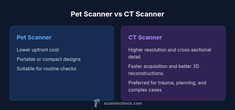
Pet scanners and CT scanners both provide cross-sectional veterinary imaging, but CT usually delivers higher resolution and faster acquisition for pets. Pet-specific or adapted scanners are more accessible and affordable, often suitable for routine checks. The choice depends on the clinical question, patient size, and facility capabilities; CT is preferred for trauma, surgical planning, and detailed anatomy.
Understanding the core distinction between a pet scanner and CT for animals
In veterinary imaging, the question pet owners and clinicians often ask is whether a dedicated pet scanner, a general-purpose imaging device adapted for animals, or a full CT (computed tomography) system best fits the clinical scenario. The decision hinges on the diagnostic goal, the patient’s size and temperament, and the clinic’s access to technology. Scanner Check emphasizes that the term pet scanner can refer to several configurations, from compact, veterinary-tailored units to human scanners repurposed for animal patients. The CT scanner, by contrast, provides cross‑sectional imaging with precise grayscale data, making it especially powerful when subtle anatomy and spatial relationships matter. For the typical pet, understanding these distinctions helps clinicians tailor imaging plans that maximize diagnostic yield while minimizing stress and radiation exposure.
How imaging hardware translates to results in veterinary patients
The hardware behind animal imaging shapes what the clinician can see. Veterinary-known scanners must accommodate a wide range of body sizes—from small felines to large dogs—and often require adaptable positioning that minimizes motion. Pet-specific devices tend to be more compact, easier to fit into a general practice, and designed for quick setup. CT scanners use a rotating X-ray source and detectors to acquire volumetric data, enabling rapid multi‑plane reconstructions and 3D modeling. The gantry size, detector configuration, and software tools determine how effectively a clinic can capture the anatomy of interest, whether it’s the skull, spine, chest, or abdomen. In practice, this means that a CT suite often delivers more consistent cross-sectional detail across body regions, while a good pet scanner may excel in more straightforward or routine imaging tasks.
Image quality: resolution, contrast, and artifacts
Image quality is defined by resolution, contrast, and the presence of artifacts. CT generally offers higher spatial resolution and sharper bone edges, with the ability to quantify tissue density via Hounsfield units. This precision supports accurate fracture assessment, integration with surgical planning, and detailed evaluation of complex thoracic or abdominal structures. Pet scanners, while improving, usually trade some resolution for practicality and cost. They are often optimized for routine screenings or targeted views, which can be perfectly adequate for common conditions but may miss subtle lesions that require finer cross‑sectional detail. Artifacts from motion, metallic implants, or patient cooperation are common considerations in both modalities, and sedations protocols commonly influence the final image quality.
Operational realities: patient handling, anesthesia, and workflow
Best practice in veterinary imaging integrates patient welfare with diagnostic goals. CT scanning frequently requires anesthesia or deep sedation to minimize motion during rapid, multi-slice acquisitions. The anesthesia plan, monitoring requirements, and post‑procedure recovery all impact workflow. Pet scanners may offer shorter procedures and simpler handling in settings lacking full anesthesia support, yet they may depend more on operator experience to optimize positioning and image interpretation. Scheduling, staff training, and the availability of radiologists or veterinary imaging specialists shape day-to-day workflow. Practitioners should weigh the need for speed and cross‑sectional detail against the resources and safety considerations of their clinic when deciding which system to rely on for a given case.
Radiation safety and dose considerations in veterinary practice
Radiation safety is central to choosing between a pet scanner and a CT system. CT delivers significant diagnostic value but comes with higher radiation exposure per exam, so dose optimization and shielding are essential. Clinics apply ALARA (as low as reasonably achievable) principles, tailor scan parameters to the smallest feasible field of view, and use dose‑modulation features when available. Pet scanners can be designed to minimize dose for routine scans, especially in smaller patients or when multiple imaging sessions are anticipated. Effective safety planning also includes operator protection, patient handling strategies to reduce repeat exposures, and clear communication with pet guardians about the necessity and benefits of imaging versus potential risks.
Cost, availability, and workflow implications
Economic considerations are a practical reality for clinics choosing between pet scanners and CT systems. Upfront purchase price, maintenance costs, service contracts, and required facility upgrades all influence total cost of ownership. CT scanners generally require more substantial investment and specialized support, but deliver broad diagnostic capability across soft tissue, bone, and vascular assessments. Pet scanners tend to have lower entry costs, shorter installation timelines, and easier integration into general practices, which can translate into quicker ROI for routine imaging. Workflow implications include appointment length, need for anesthesia, image processing time, and the potential for collaborations with centralized radiology services when more advanced imaging is required.
Clinical scenarios: when to choose CT vs pet scanner
Different clinical questions tilt the balance toward one modality or the other. For acute trauma, suspected skull or chest fractures, or oncologic staging requiring precise lesion localization and 3D planning, CT is often preferred. For routine dental checks, soft tissue screening, or follow-up imaging in small clinics, a well‑tuned pet scanner can meet the diagnostic goal efficiently. Orthopedic planning, metastatic disease evaluation, and complex organ imaging frequently benefit from the superior cross‑sectional resolution of CT, while surveillance imaging in smaller, stable patients might be well served by a pet scanner. Clinicians should assess the question, patient condition, and logistical constraints to craft an imaging plan.
Practical tips for clinics planning to acquire CT or pet scanners
When budgeting for imaging, consider your expected caseload, patient demographics, and the typical diagnostic questions your team faces. If CT is chosen, plan for room design, radiation shielding, and the need for trained staff or ongoing tele-radiology support. If a pet scanner is pursued, look for user-friendly interfaces, efficient software for 3D reconstructions, and compatibility with the clinic’s anesthesia and monitoring workflows. Training, maintenance contracts, and data management strategies (PACS and cloud storage) will also influence uptime and throughput. Finally, keep an eye on emerging AI-assisted tools that can aid with segmentation, measurement, and lesion characterization, potentially narrowing the gap in image interpretation between modalities.
Emerging trends and future directions in veterinary imaging
The field is evolving toward shorter, safer scans, improved intraoperative imaging, and integrated AI for rapid interpretation. Portable or compact CT solutions are expanding access in general practices, while dedicated veterinary scanners continue to push higher resolutions and faster throughput. Cross‑disciplinary collaborations with human imaging experts and participation in continuing education help clinics stay current. As data science advances, clinicians may rely more on automated lesion detection, workflow optimization, and enhanced dose tracking, enabling better diagnostic decisions without compromising patient well‑being.
Comparison
| Feature | Pet scanner | CT scanner |
|---|---|---|
| Imaging modality basics | Dedicated veterinary imaging or adapted devices | Cross-sectional imaging using computed tomography |
| Image resolution | Lower to moderate; device-dependent | Higher resolution with precise bone detail |
| Acquisition speed | Longer per study; sometimes motion-limited | Very fast per slice with reduced motion artifacts |
| Anatomical coverage | Often limited to targeted views or small volumes | Full-body or organ-specific coverage with 3D reconstructions |
| Radiation dose and safety | Lower dose in some configurations | Higher potential dose with dose management strategies |
| Portability and access | Often portable or compact; fits smaller clinics | Typically fixed in imaging suite; limited portability |
| Best for | Routine checks and quick-turnaround cases | Trauma, complex anatomy, surgical planning |
Pros
- Lower equipment cost and simpler maintenance
- Easier integration into general practice
- Quicker setup for routine assessments in small clinics
- Lower overall staff training demands compared to CT setup
Drawbacks
- Lower resolution and limited cross-sectional detail
- Reduced capabilities for complex anatomy or trauma planning
- Less consistent performance across a wide range of body sizes
- Potentially longer exam times for comprehensive assessments
CT scanner generally provides superior diagnostic detail for most veterinary cross‑sectional imaging; pet scanners offer practical value where access and budget are limited.
Use CT for trauma, oncology staging, and detailed surgical planning. Consider a pet scanner when you need routine imaging with lower upfront costs and easier workflow. The Scanner Check team emphasizes matching modality to the clinical question and clinic capabilities.
Common Questions
What is the difference between a pet scanner and a CT scanner in veterinary imaging?
A pet scanner typically refers to veterinary-tailored devices or adapted human scanners used for routine imaging, offering easier access and lower costs. A CT scanner provides detailed cross-sectional images with higher resolution and the ability to reconstruct 3D views, which is particularly useful for complex anatomy and surgical planning.
Pet scanners are more affordable and easier to use day-to-day, while CT gives sharper, 3D‑ready images for complicated cases.
When is a pet scanner sufficient for veterinary care?
For routine screening, dental imaging, and basic anatomical assessments where extreme detail is not required, a pet scanner can meet diagnostic needs effectively. In such cases, workflow efficiency and patient comfort often drive the decision.
For simple, routine checks, a pet scanner often does the job well.
Can a CT scanner be used for soft tissue imaging in pets?
Yes, CT is excellent for many soft tissue assessments, especially with contrast enhancement. It is particularly valuable when detailed cross-sectional visualization is needed, though MRI may provide superior soft tissue contrast for certain indications.
CT works great for soft tissues with contrast; MRI can be better for some soft-tissue details.
What are the cost implications between pet scanners and CT scanners?
CT systems typically involve higher upfront costs, installation, and ongoing maintenance, but offer broad diagnostic capability. Pet scanners tend to be cheaper to acquire and run, making them attractive for clinics with budget constraints and a focus on routine imaging.
CT costs more upfront, but pet scanners save money and space for routine imaging.
Is radiation exposure a concern in veterinary imaging with CT?
Yes, radiation exposure matters. Clinics implement dose optimization, shielding, and protective protocols to minimize exposure for patients, staff, and guardians. ALARA principles guide imaging decisions across both modalities.
Radiation safety is important; clinics optimize doses and protect everyone involved.
Are there alternatives to CT for diagnostic imaging in pets?
Yes. Ultrasound and X-ray radiography are common alternatives, with MRI used for certain soft-tissue indications. Each modality has its own strengths and is chosen based on the clinical question and available resources.
There are other tools like ultrasound and MRI; CT remains special for detailed cross-sections.
Key Takeaways
- Define the clinical question first to pick the right modality
- CT delivers higher resolution and cross-sectional detail
- Pet scanners provide cost-effective, accessible imaging for routine cases
- Plan for anesthesia, safety, and workflow when acquiring new equipment
- Stay informed on AI aids and dose-optimization techniques