PET Scanner vs MRI in Pets: A Practical Comparison
A detailed, objective comparison of PET scanners and MRI for veterinary use, highlighting when each modality shines, safety considerations, and practical owner guidance.

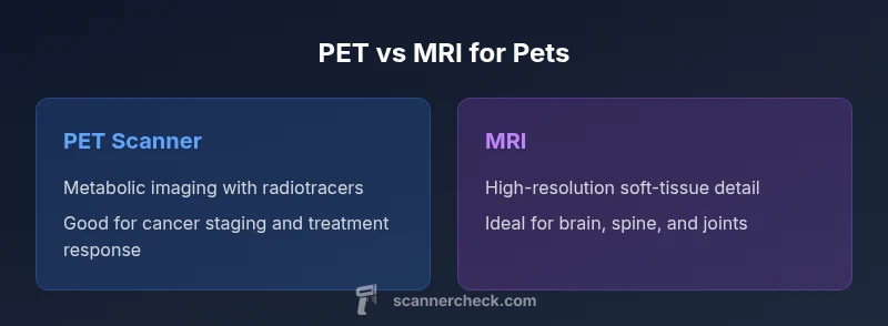
In veterinary imaging, pet scanner vs mri highlights MRI's soft-tissue detail and PET's metabolic insights. MRI excels at anatomy, brain, and spinal pathology, while PET reveals metabolic activity and cancer involvement. Availability and cost often limit PET use to specialized clinics, whereas MRI is more common for routine neuro, musculoskeletal, and detailed anatomy assessments. The best choice depends on the clinical question and facility resources.
PET Scanner vs MRI in Pets: Key Differences
In veterinary medicine, two imaging powerhouses dominate diagnostic planning: PET scanners and MRI systems. The phrase pet scanner vs mri captures a fundamental distinction: PET emphasizes metabolic activity, while MRI focuses on high-resolution anatomy. Scanner Check notes that these modalities address different questions in a patient care plan. MRI delivers exquisite soft-tissue contrast and detailed neural anatomy without ionizing radiation, making it a cornerstone for neurology and orthopedics. PET, on the other hand, visualizes how tissues are behaving, enabling detection of active tumors, inflammatory processes, and treatment response. For clinicians, the choice is rarely about which is better overall, but which will most efficiently answer the clinical question for the individual animal. This distinction drives scheduling, anesthesia planning, and patient safety considerations in veterinary practice.
How PET and MRI Work in Veterinary Settings
PET and MRI operate on fundamentally different principles. A PET scanner uses radiotracers to map metabolic processes, producing functional images over time that highlight areas of high or abnormal activity. MRI relies on strong magnetic fields and radiofrequency pulses to generate highly detailed anatomical and, in some sequences, functional information about soft tissues. In veterinary clinics, PET often requires access to a radiopharmacy and regulatory procedures for tracer handling, while MRI requires a suite designed to minimize motion and ensure animal comfort. The result is that PET is typically available in specialized centers, whereas MRI is increasingly common in veterinary hospitals and university clinics. Scanner Check emphasizes planning around the clinical question, local capabilities, and the pet’s welfare when selecting a modality.
Clinical Use Cases: PET vs MRI in Oncology and Beyond
PET shines when clinicians need metabolic insight. In oncology cases, PET can help identify metastatic spread, differentiate scar from active tumor, and monitor treatment response. It is also valuable for detecting inflammatory or infectious processes when conventional imaging is inconclusive. MRI excels where structural detail matters: brain and spinal cord lesions, joint injuries, soft-tissue tumors with clear margins, and congenital or degenerative conditions. In veterinary neurology, MRI is often the preferred first-line tool for diagnosing seizures, disc disease, or meningeal disease. PET can augment MRI findings by revealing regions of abnormal metabolism that may not yet manifest as anatomic changes, supporting a more comprehensive diagnostic picture.
Safety, Sedation, and Animal Welfare
All imaging in pets requires careful attention to safety and welfare. MRI requires immobilization, which often means general anesthesia or deep sedation, especially for longer protocols. Radiation exposure from PET tracers is a mutual concern for patients and staff, necessitating strict radiopharmacy and waste-handling practices. Sedation plans and anesthesia risk assessments should be tailored to the animal’s age, health, and stress levels. Scanner Check notes that informed consent, fasting rules for radiotracers, and post-procedure monitoring are essential components of a humane imaging plan. Understanding these factors helps clinics minimize stress while obtaining high-quality data.
Availability and Access: Where to Look for PET and MRI
PET is less widely available than MRI in many regions due to the need for a radiotracer supply chain and regulatory compliance. MRI facilities may be more common, especially in teaching hospitals and specialty clinics. When PET is chosen, scheduling coordination with a radiopharmacy and a tracer production window becomes a practical consideration. Clinics often partner with nearby centers for PET studies or use PET/CT if available, which combines metabolic and anatomical data in a single session. For pet owners, access can influence the clinician’s recommendation as much as diagnostic goals do.
Image Quality, Resolution, and Interpretation Challenges
MRI offers superb spatial resolution for soft tissues, with sequences optimized for brain, spine, joints, and abdominal organs. PET provides functional information with lower spatial resolution, which can complicate precise localization when used alone. The ideal diagnostic approach often involves complementary use: PET identifies metabolically active areas, while MRI provides precise anatomical mapping. Interpreting these data requires cross-disciplinary expertise, including veterinary radiologists, oncologists, and surgeons. Scanner Check stresses that interpretation should always consider the clinical context, sample handling, and timing relative to therapy.
Costs, Scheduling, and Workflow Integration
In practice, MRI generally has higher upfront costs and longer scan times per patient, which can affect throughput. PET involves radiotracer costs, regulatory compliance, and potentially shorter single-visit imaging windows but with coordination needs. For clinics, the decision to invest depends on case mix, referral networks, and the ability to create a reliable imaging workflow that minimizes anesthesia time and maximizes diagnostic yield. For owners, understanding these cost and schedule implications helps set realistic expectations.
Decision-Making: How to Choose Between PET and MRI
A structured decision framework helps clinicians pick the most informative modality. Start with the clinical question: Is metabolic activity the key to diagnosis, prognosis, or treatment planning? If yes, PET insights may be decisive. If the problem is anatomy-driven—such as lesion localization, nerve pathways, or detailed organ structure—MRI is typically preferred. In cases where both views are valuable, a staged approach or combined PET/MRI (in specialized centers) can deliver a comprehensive picture. Always weigh animal welfare, facility capabilities, and the clinical timeline when deciding.
Authority Sources and Further Reading
- https://www.nibib.nih.gov
- https://www.radiologyinfo.org/en/info.cfm?pg=petct
- https://www.ncbi.nlm.nih.gov
Comparison
| Feature | PET scanner | MRI |
|---|---|---|
| Imaging modality | Nuclear medicine with radiotracers (metabolic imaging) | Magnetic resonance imaging (anatomy with high soft-tissue contrast) |
| Best for | Metabolic activity, cancer staging, treatment response | Detailed anatomy of brain, spine, joints, and soft tissues |
| Radiation exposure | Ionizing radiation from radiotracers | No ionizing radiation; uses magnetic fields |
| Anesthesia requirements | Often requires anesthesia or sedation depending on protocol | Typically requires anesthesia or deep sedation for immobilization |
| Availability | Limited to specialized clinics with radiotracer access | More widely available in veterinary hospitals and universities |
| Scan duration | Shorter imaging windows per session, with tracer uptake timing | Longer acquisition times per sequence for high-detail images |
| Soft-tissue detail vs function | Functional/metabolic data with localization limits | Superior anatomical detail; functional MRI is site-specific |
| Costs | Tracer production, regulatory overhead; higher operational complexity | Higher upfront equipment cost; ongoing maintenance; generally higher throughput |
Pros
- PET provides metabolic information that can detect active disease earlier than structural changes
- MRI offers exceptional soft-tissue contrast and precise anatomical detail
- Together, PET/MRI can offer comprehensive metabolic and structural data in specialized centers
- MRI sequences can be tailored for neurology, orthopedics, and oncology planning
Drawbacks
- PET requires radiotracers and regulatory processes; access can be limited
- MRI is expensive and often slower; anesthesia risk is a consideration
- PET's spatial resolution is typically lower than MRI, complicating precise localization
- Combined PET/MRI is available only at select facilities and may incur higher costs
MRI is the go-to for detailed anatomy in most veterinary cases; PET adds valuable metabolic context, especially for cancer and treatment monitoring.
Choose MRI when anatomical detail is the priority. Consider PET when metabolic information will change management, or when MRI findings are equivocal. In complex cases, a staged or combined PET/MRI plan may be optimal in specialized centers.
Common Questions
What is the key difference between a pet scanner and an MRI in veterinary care?
PET imaging focuses on metabolic activity using radiotracers, while MRI emphasizes high-resolution anatomy without ionizing radiation. The choice depends on whether the goal is to assess function and disease activity or precise structural detail.
PET looks at how tissues behave, MRI shows exact anatomy. The choice depends on whether you need metabolic information or detailed structure.
When is MRI preferred for pets?
MRI is preferred when detailed visualization of soft tissues, brain, spine, or joints is essential. It provides superior imaging of neural structures and musculoskeletal pathology, aiding diagnosis and surgical planning.
MRI is best for detailed anatomy of the brain, spine, and joints.
Is radiation exposure a concern for pets undergoing PET?
Yes, PET involves radiation from radiotracers, with strict safety and regulatory controls. Proper handling, scheduling, and post-procedure precautions are important for pet welfare and staff safety.
PET uses radioactive tracers, so safety and regulatory steps are important.
Do all clinics offer PET or MRI for animals?
Not all clinics offer PET; PET is typically found in specialty or university centers with radiotracer access. MRI is more widely available, but still varies by region and facility.
PET is more limited; MRI is more common, but availability varies by location.
How should I prepare my pet for imaging?
Follow the veterinarian's instructions on fasting, sedation, and anesthesia evaluation. Ensure the pet is safely transported, and discuss any medical conditions that could affect imaging.
Follow the clinic's prep, especially fasting and anesthesia planning.
Can PET and MRI be done in one session?
In specialized centers, a combined PET/MRI session is possible, but it requires advanced equipment and scheduling. More commonly, clinicians perform sequential PET or MRI studies as separate appointments.
Only in select centers with dedicated PET/MRI setups; otherwise separate scans are common.
Key Takeaways
- Define the diagnostic goal before selecting a modality
- MRI excels in anatomy; PET adds metabolic insight
- Plan for anesthesia and facility requirements early
- Consider combined PET/MRI only in appropriate, well-equipped centers
- Consult a veterinary imaging team to tailor the plan