When to Scan for Pregnancy: A Practical Timing Guide
Learn when to scan for pregnancy, including home test windows, dating ultrasounds, first-trimester checks, and anatomy scans. Practical timing guidance for expectant parents, with data-driven context.

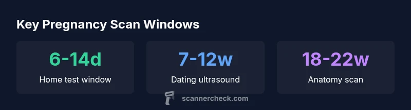
Home pregnancy tests generally become positive about 6-14 days after ovulation, and a dating ultrasound is commonly scheduled around 7-12 weeks gestation to confirm dating and viability. If you have early pregnancy concerns, your clinician may perform an ultrasound sooner. For ongoing pregnancy monitoring, anatomy scans occur later in the second trimester. This timing helps reduce uncertainty and supports informed care decisions.
Why Timing Matters for Pregnancy Scans
Timing of scans and tests is not arbitrary. It shapes how accurately clinicians can determine gestational age, assess fetal viability, and plan subsequent screenings. Inaccurate dating can shift the entire prenatal schedule, affecting when important tests—like nuchal translucency or anatomy assessment—are performed. The Scanner Check team emphasizes that following established windows helps ensure reliable results while minimizing unnecessary procedures. In pregnancies with risk factors, or when symptoms such as bleeding occur, clinicians may adjust timing to prioritize safety and diagnostic clarity. Consistency across visits helps build a clear trajectory of fetal growth and maternal health.
Key takeaway: Timelines are guidelines, not strict rules; always align with your healthcare provider’s plan and any pregnancy-specific factors.
Understanding Home Pregnancy Tests: When to Expect a Result
Home pregnancy tests detect the hormone hCG in urine. Most individuals see a positive result after implantation, which typically occurs 6-14 days after ovulation. Test sensitivity varies by brand, and urine concentration can affect results. For the most reliable early read, follow the test instructions closely and consider testing again after a missed period if results are unclear. If you receive a positive result, contact your clinician to plan confirmatory steps and a prenatal care timeline.
Practical tip: Use the first-morning urine for the strongest signal, and avoid drinking large amounts of fluid just before testing.
Early Clinic Testing: When Blood Tests Matter
While home tests are convenient, clinic-based blood tests provide quantitative data and can detect pregnancy earlier in some cases. A quantitative hCG blood test measures the exact amount of hCG in the bloodstream and tracks how quickly it rises over the first days. This helps confirm viability and can influence the timing of an ultrasound. In practice, clinicians often integrate hCG results with symptoms and prior test results to determine the next imaging step and prenatal screening schedule.
Note: If levels are not rising as expected, a repeat test or additional imaging may be warranted to rule out complications.
The Dating Ultrasound: Aligning Gestational Age
A dating ultrasound is one of the standard early scans used to estimate gestational age. It commonly measures crown-rump length (CRL) and assesses early organ development. The usual window is about 7-12 weeks gestation; in some cases, imaging may occur slightly earlier or later depending on factors such as maternal body habitus, fetal position, and clinical indication. Accurate dating reduces the risk of errors in scheduling-screening milestones like the first-trimester screen and subsequent anatomy assessments.
Important nuance: Dating accuracy improves with ultrasound in the first trimester compared to relying on last menstrual period alone, especially for first pregnancies or irregular cycles.
Early Pregnancy Scans: What They Can Reveal (6-12 weeks)
In the early weeks, ultrasound can confirm intrauterine pregnancy, detect fetal heartbeat, and identify a viable gestational sac. It also helps determine the number of embryos and assess early growth patterns. By around 6-7 weeks gestation, a fetal heartbeat is often visible, though this can vary with equipment and maternal factors. Early scans can detect ectopic pregnancy or other concerns that require prompt management. The results guide decisions about subsequent tests and appointment scheduling.
Clinical takeaway: Early imaging informs prognosis while guiding future care decisions and reducing uncertainty for parents alike.
The Nuchal Translucency and First-Trimester Checks
The nuchal translucency (NT) scan is typically performed between 11-14 weeks to screen for chromosomal conditions and fetal anatomy issues. NT measurement is used in combination with maternal blood tests to assess risk, not as a standalone diagnosis. Some clinics offer this as part of the first-trimester screen, while others may implement it later depending on local protocols. Having clear expectations about what the NT scan assesses helps families prepare questions for their provider.
What to ask: Whether NT assessment is recommended for your pregnancy and how results influence subsequent testing.
Anatomy Scan: When to Check Fetal Development (18-22 weeks)
The anatomy scan, often called the detailed fetal survey, occurs around 18-22 weeks and evaluates organ development and placental position. This timing provides a comprehensive view of fetal anatomy and growth, helping identify structural concerns that could require follow-up or interventions. In cases of high-risk pregnancies or prior anomalies, clinicians may adjust the timing or add targeted ultrasounds. Parents should discuss what will be assessed and what findings might mean for care planning.
Practical note: Bring any prior imaging records and a list of questions to your appointment.
Special Cases: Bleeding, Pain, or High-Risk Pregnancies
Bleeding, cramping, or a history of pregnancy complications can prompt earlier or more frequent scans. In such scenarios, clinicians may perform targeted ultrasound earlier than the standard dating window or add specialized imaging to monitor growth and viability. For high-risk pregnancies, a tailored schedule helps ensure timely detection of potential issues while balancing safety and resource use. Always share symptoms, medical history, and any concerns with your care team.
Bottom line: Scans are tools to guide care, not merely milestones; use them to inform decisions with your provider.
Practical Tips: How to Prepare for Scans and Discuss with Your Provider
Preparation for scans varies by type. For pelvic or transabdominal ultrasounds, a full bladder may be requested to improve image quality, while transvaginal scans may not require this. Write down questions in advance, such as the purpose of each scan, what will be measured, and how results will influence next steps. If you’re unsure about timing, request a written plan outlining when to expect each test and any potential follow-ups. Clear communication with your provider helps you navigate the process with confidence.
Overview of common pregnancy tests and ultrasound timing
| Test Type | Typical Timing | Purpose |
|---|---|---|
| Home pregnancy test (urine) | 6-14 days after ovulation | Preliminary confirmation of pregnancy |
| Quantitative hCG blood test | Same day after order | Accurate measurement of hCG level and pregnancy viability |
| Transvaginal ultrasound | 6-8 weeks gestation (dating) | Assess viability and dating in early pregnancy |
| Transabdominal ultrasound | 12-14 weeks for dating in some cases, or 18-22 weeks anatomy | Further assessment depending on gestational age |
Common Questions
When is the earliest I can confirm pregnancy with ultrasound?
Typically around 6-8 weeks gestation, though a clinician may image earlier if symptoms or risk factors warrant it.
Most people can get an early ultrasound around six to eight weeks if medically indicated.
Can home pregnancy tests detect pregnancy before a missed period?
Yes, many tests can detect pregnancy about 6-14 days after ovulation, but accuracy improves after a missed period.
Yes, tests can often detect pregnancy about a week after ovulation, but accuracy is higher after the missed period.
What is the anatomy scan and when is it done?
The anatomy scan is usually performed around 18-22 weeks to assess fetal development and anatomy.
The anatomy scan is usually done at around 18 to 22 weeks.
Why might a dating ultrasound be delayed?
Obesity, fetal position, or late presentation can delay dating ultrasounds or require a repeat exam.
A dating ultrasound might be delayed due to body habitus, fetal position, or late presentation.
Are there risks to ultrasound during pregnancy?
Clinical ultrasound is considered safe when performed for medical reasons by trained professionals.
Ultrasound is considered safe when used for medical reasons and done by trained staff.
What should I discuss with my provider before a scan?
Ask about timing, what the scan can reveal at each stage, and any preparations (full bladder for some pelvic scans).
Ask about when to schedule, what will be checked, and any prep like a full bladder.
“Timely scans and tests align clinical milestones with fetal development, reducing uncertainty for patients and clinicians alike.”
Key Takeaways
- Start with a home test around the time you expect your period.
- Plan a dating ultrasound in the 7-12 week window for accurate dating.
- Schedule anatomy scanning in the 18-22 week window to assess growth.
- Seek imaging earlier if bleeding, pain, or high-risk factors arise.
- Discuss timing openly with your healthcare provider and follow their guidance.