Difference Between a Scan and an MRI: What You Need to Know
Explore the key differences between a medical scan and an MRI, including how they work, safety, costs, and when each is preferred. Clear guidance for patients, clinicians, and tech enthusiasts.

In medical imaging, a 'scan' is a broad term for any imaging exam, while an MRI is a specific modality that uses strong magnets and radio waves to image soft tissues. The difference between a scan and an MRI lies in the technology, safety profile, timing, and typical clinical use. This guide highlights the core contrasts to help patients and clinicians choose the right test.
Terminology and scope
In everyday clinical language, people often say they are getting a scan when they mean an imaging test of the body. However, the word scan is a broad umbrella that covers several imaging technologies, including X-ray, computed tomography (CT), ultrasound, nuclear medicine, and MRI. An MRI, by contrast, is a specific modality that relies on strong magnetic fields and radiofrequency pulses to create detailed images of soft tissues such as the brain, spinal cord, ligaments, and organs. Understanding this distinction is essential when a clinician orders tests, because the information each modality yields and the constraints it imposes can be markedly different. According to Scanner Check, clarifying the modality up front can reduce confusion and improve planning for subsequent steps in care.
How MRI works
MRI uses a powerful magnetic field to align protons in the body, then radiofrequency pulses perturb that alignment. As protons realign, they emit signals that a computer converts into cross-sectional images. Different MRI sequences emphasize various tissues—fat, water, and the presence of blood or scar—providing exceptional soft-tissue contrast. Because MRI does not rely on ionizing radiation, it is particularly valued for brain and spinal imaging, joint evaluations, and certain abdominal and pelvic studies. The scanning process includes several steps: positioning, calibration, sequence selection, and sometimes the use of contrast agents to enhance visibility of specific structures. The result is high-resolution images that help detect subtle abnormalities.
What a generic 'scan' can include
When most people hear "scan," they might think of a CT scan or a traditional X-ray. CT uses X-ray beams and detectors to generate cross-sectional images quickly and is excellent for bone detail and acute injuries. Ultrasound uses sound waves to create real-time images and is invaluable for fetal assessments and abdominal organs in many settings. Nuclear medicine scans involve adding radioactive tracers to assess organ function. In contrast to MRI, these modalities rely on different physical principles and deliver distinct kinds of information. Understanding that the term scan can refer to multiple technologies helps set expectations for accuracy, safety, and what doctors can infer about pathology.
Radiation exposure and safety implications
A major differentiator is radiation. MRI emits no ionizing radiation and is generally considered safe for many patients, though there are safety rules around implants and devices. CT and X-ray, on the other hand, involve ionizing radiation, which carries a small, cumulative risk with repeated exposure. When choosing between a scan and an MRI, clinicians weigh the diagnostic benefit against radiation exposure, especially in pediatric patients or when multiple follow-up tests are anticipated. Gadolinium-based contrast agents used in MRI can carry rare risks in susceptible individuals, while iodinated contrast used in CT may trigger allergies or kidney concerns in some patients. Scanner Check emphasizes patient-specific risk assessment as a central part of the decision.
Image quality, resolution, and what each test excels at
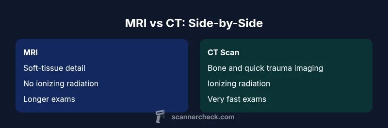
MRI tends to excel at soft-tissue contrast, enabling detection of subtle lesions in the brain, spinal cord, joints, and organs. CT provides rapid, high-resolution images of bone and acute injuries, as well as chest and abdominal scans that require quick decision-making. Scan quality is influenced by patient factors (movement, implants), machine capabilities, and protocol choices. In practice, clinicians may start with a CT for an emergency evaluation and then use MRI for detailed soft-tissue assessment. The choice hinges on the clinical question: Is the priority bone detail, overall anatomy, or soft-tissue pathology?
Patient experience and comfort considerations
MRI scanners are large, noisy, and magnet-friendly environments; some patients feel claustrophobic, and tall or anxious patients may require sedation or an open MRI option. CT scans are typically shorter in duration and may feel less intimidating but expose patients to ionizing radiation. The patient experience also differs in terms of preparation: MRI often requires removing metal, reviewing implants, and sometimes fasting if contrast is planned, while CT may have different preparation guidelines depending on contrast use. Clinicians tailor the approach to minimize discomfort while maximizing diagnostic yield. Scanner Check notes that comfort, safety, and clear communication with the patient are foundational to successful imaging.
Preparation, contraindications, and safety steps
Before MRI, patients submit a detailed safety questionnaire about implants, pacemakers, or metal fragments. Certain implants are MRI-compatible, but some may not be, necessitating alternative imaging. Clothing with removable metal, dentures, and jewelry are typically removed. For CT with contrast, kidney function and hydration status are assessed, and patients may be advised to fast. CT has fewer physical constraints than MRI but involves radiation exposure. In any case, disclose prior imaging, allergies, and pregnancy status, as these factors influence the test choice and contrast decisions.
Clinical decision pathways: when to use MRI versus a general scan
The decision to order an MRI versus a general scan often centers on the suspected pathology. MRI is preferred for soft-tissue assessment, neurological conditions, and musculoskeletal injuries where detail matters. CT is often the go-to in acute trauma, chest imaging, or when rapid results are needed. In some cases, a combination of scans provides the most comprehensive view. Clinicians use guidelines, patient history, and physical examination findings to determine which modality will answer the clinical question most efficiently and safely.
Costs, turnaround, and accessibility considerations
Accessibility and cost vary by region and facility. MRI tends to be more expensive and less available on short notice, which can impact scheduling and patient cost. CT is widely available, faster, and commonly used in emergency departments for rapid decision-making. Insurance coverage, facility capabilities, and local protocols influence the actual cost and waiting time. Scanner Check highlights the importance of understanding cost implications and potential need for follow-up imaging when planning care.
Practical tips for patients and clinicians when planning imaging
Engage in a pre-test discussion about the exact clinical question, expected image quality, and possible need for contrast. If time allows, consider MRI for better soft-tissue detail and CT for quick assessment. Prepare by asking about safety, required sedation, or contrast, and what to expect during the procedure. After the test, request a clear report focusing on the clinical implications rather than just the radiology findings. The goal is a test that meaningfully informs treatment decisions.
Quick reference: choosing the right test in common scenarios
In head trauma, CT is often the first step due to speed and bone detail, followed by MRI if soft-tissue injury remains uncertain. In suspected spinal cord pathology or joint injuries, MRI may reveal subtle edema or ligament tears not visible on CT. In abdominal or pelvic interrogation, the choice depends on suspected pathology and the need for functional information versus anatomic detail. This section summarizes practical decision cues for clinicians and patients alike.
Comparison
| Feature | MRI | CT scan |
|---|---|---|
| Imaging modality and principle | Magnetic fields + radio waves; excellent soft-tissue contrast | Ionizing radiation imaging using X-rays; fast and bone-focused |
| Radiation exposure | none or negligible with standard protocols | ionizing radiation; exposure varies by protocol |
| Typical use cases | Soft-tissue detail (brain, spine, joints, organs) | Acute trauma, bone injuries, chest/abdominal screening |
| Scan duration | Typically 20-60 minutes depending on protocol | A few minutes for basic CT; longer with contrast |
| Contraindications/risks | Metal implants, claustrophobia, rare contrast risks | Allergy to iodinated contrast; kidney considerations; pregnancy considerations |
| Contrast agents | Gadolinium-based agents (when used) or none | Iodinated contrast; risk of allergy or nephrotoxicity |
| Image artifacts | Movement, implants; slower sequences can be affected | Metal artifacts; motion can degrade image quality but is often manageable |
| Cost/availability | Higher cost; longer scheduling in some regions | Faster, widely available; lower per-scan cost in many settings |
Pros
- MRI provides superior soft-tissue contrast without ionizing radiation
- CT scans are fast, widely available, and highly effective for bone and emergency imaging
- MRI offers advanced imaging options in some centers (e.g., functional sequences)
Drawbacks
- MRI can be time-consuming and claustrophobic for some patients
- CT involves ionizing radiation and carries cumulative exposure risk
- MRI may be more expensive and less accessible in some regions
MRI is the preferred test for soft-tissue detail when safe and available; CT or other scans are often better for fast, bone-focused assessments.
Choose MRI when detailed soft-tissue information is crucial and there are no contraindications. Opt for a CT or other scan when speed, bone visualization, or accessibility are the priority, or when radiation exposure must be minimized in sensitive populations.
Common Questions
What is the difference between a scan and an MRI?
A scan is a broad term for any medical imaging test (CT, MRI, X-ray, ultrasound). MRI is a specific imaging modality that uses magnets and radio waves to image soft tissues and does not involve ionizing radiation. The two differ in technology, safety, and typical clinical use.
A scan refers to any imaging test, while MRI is a specific technology that uses magnets; MRI avoids radiation and is best for soft tissue. If you’re unsure which test you need, ask your clinician to clarify the modality and purpose.
Is MRI safe for people with metal implants?
Many implants are MRI-conditional, but some are not. Safety depends on the implant type, location, and device. Always inform the imaging team about any implants to determine whether an MRI is appropriate.
Tell the tech about every implant or device you have so they can assess MRI safety.
Does a scan always involve radiation?
Not all scans involve radiation. MRI does not use ionizing radiation, while CT and some X-ray exams do. The choice of test balances information gained against radiation exposure.
MRI usually has no radiation; CT and X-ray tests do involve it.
How long does an MRI exam take?
An MRI exam typically takes 20 to 60 minutes, depending on the body area and the number of sequences. Some studies may be shorter, while detailed protocols can extend the time.
Most MRIs run under an hour, but it can be longer for thorough scans.
Can MRI images show bone fractures as well as soft tissues?
MRI can image bone and soft tissue, but CT is generally superior for detecting acute fractures and fine bone detail. MRI’s strength lies in soft-tissue and marrow assessment.
MRI can show bone issues, but CT is typically better for fractures.
What should I ask my doctor before a scan or MRI?
Ask about the exact modality, reason for the test, contrast needs, duration, safety considerations, and how the results will influence treatment decisions. Clarify any risks you're concerned about.
Ask which test is best for your condition and what to expect during the procedure.
Key Takeaways
- Understand that a 'scan' is a broad term; MRI is a specific modality.
- MRI uses magnets and radio waves; CT uses X-ray radiation.
- MRI avoids ionizing radiation; CT does not.
- Test choice depends on anatomy, urgency, and safety considerations.
- Discuss contrast needs and potential contraindications with your clinician.